Ultrasound-Guided Nerve Block in Breast Surgery
PECS block type 1 is an ultrasound-guided interfacial block that can be used to manage postoperative analgesia after breast surgery. A local anesthetic solution is injected into the interfacial area between the Pectoralis Major (PMm) and Pectoralis Minor (PMn) muscles during the procedure (Pmm).
Breast surgery has undergone changes in recent years, encouraging new initiatives for the anesthetic management of these patients in order to achieve maximum quality and rapid recovery. The fundamental tool that has allowed a significant improvement in the progress of regional anesthesia for breast disease has been ultrasound, boosting the description and introduction into clinical practice of interfascial chest wall blocks, although the reference standard is still the paravertebral block. It is very likely that these blocks will change the protocols in the coming years.
Ultrasound has traditionally been within the purview of radiologists, but surgeons are finding it an effective tool they can use to locate occult lesions in the breast and to differentiate between benign and malignant.
Outpatient-based ultrasound performed by surgeons frees up the resources of radiology departments, allowing them to focus upon patients requiring more complex diagnostic and interventional procedures. For surgeons to competently perform intra-operative and outpatient-based ultrasounds, a period of formal ultrasound training is necessary to acquire knowledge of ultrasound skills and techniques.
Ultrasound imaging of the breast uses sound waves to produce pictures of the internal structures of the breast. It is used to help diagnose breast lumps or other abnormalities found during a physical exam or on a mammogram or breast MRI. Ultrasound is safe, non-invasive, and does not use radiation.
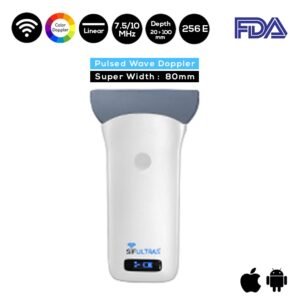
SIFULTRAS-5.39 is a special ultrasound technique that evaluates the movement of materials in the body. It allows the doctor to see and evaluate the breast. Ultrasound of the breast produces a picture of the internal structures of the breast.
SIFULTRAS-5.39 is a super-width ultrasound scanner with 256 elements. As a result, provides excellent resolution. Furthermore, the probe has a frequency range of 7.5 – 10 MHz and a visual depth of up to 100mm. The SIFULTRAS-5.39 is suitable for breast and hip bone inspections.
The fundamental tool that has allowed a significant improvement in the progress of regional anesthesia for breast disease has been ultrasound, boosting the description and introduction into clinical practice
References: Ultrasound-guided pectoral nerve block for pain control after breast augmentation