Ejection Fraction is a measurement that doctors use to calculate the percentage of blood flowing out of the ventricles ( the right and left lower chambers in the heart that enable it to pump blood into the body) with each contraction.

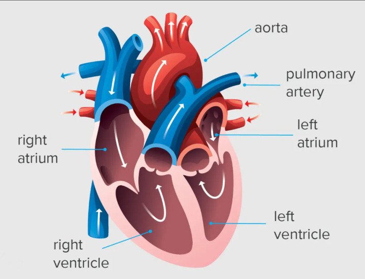
Ultrasound scanner-guided Ejection Fraction measurement.

 Ejection Fraction is a measurement that doctors use to calculate the percentage of blood flowing out of the ventricles ( the right and left lower chambers in the heart that enable it to pump blood into the body) with each contraction. Which Ultrasound Scanner is the best for Ejection Fraction

Read More »
Infertility Ultrasound

Infertility Ultrasound

Infertility ultrasound is preferred and usually mandatory for women who are having difficulty conceiving. In order to identify the abnormality that is causing this problem, the obstetricians and gynecologists (OB-GYNs) use an ultrasound probe to observe and assess female abdominal and pelvic structures starting from day 3 of the cycle.  In order to

Read More »
musculosketal-ultrasound-1

Musculoskeletal Ultrasound

Musculoskeletal ultrasound is used to allow physicians to effectively visualize and detect sprains, strains, tears, trapped nerves, arthritis, and other musculoskeletal conditions and efficiently treat them. This procedure does not require special preparation. Leaving jewelry at home and wearing loose, comfortable clothing is enough to undergo it. Which device is the most suitable

Read More »
Bladder_anatomy

Bladder Ultrasound

The bladder is an organ made of smooth muscle. It stores urine until it’s released when you go to the bathroom. The most common reason for bladder ultrasound is to check bladder draining. The urine that remains in the bladder after urinating (“post void residual”) is measured. If urine remains,

Read More »
Ultrasound examination is specific for non-invasive detection of congenital heart disease, rheumatic heart disease, and myxopathy of the heart

The Significance of Doppler Ultrasound Examination

Because ultrasound has the advantages of flexible operation, simple, no radiation, and affordability, it has been widely used in clinics. It is currently the preferred method for liver, spleen, gallbladder, pancreas, urinary system, gynecology, blood vessels, fetal examination… Even, very small lesions can be found on ultrasound. Interventional ultrasound can also

Read More »
Carotid_artery_stenosis

Carotid Artery Stenosis Ultrasound Diagnosis

Carotid artery stenosis (CAS), atherosclerotic narrowing of the extracranial carotid arteries, is a risk factor for ischemic stroke. Which makes it clinically significant. The clinical finding of a carotid bruit on auscultation is non-specfic; therefore, hemodynamically significant CAS cannot be determined by physical examination alone Of the imaging modalities available

Read More »
hydrosalpinx

Hydrosalpinx Ultrasound

Hydrosalpinx is a descriptive term and refers to fluid-filled dilatation of the fallopian tube. If the fluid id infected, i.e. pus, then it a pyosalpinx, if bloody, then hematosalpinx. Infertility due to fallopian tube factors occupies the first place among female infertility. In recent years, there has been an increase

Read More »
pediatric ultrasound

Advantages of Pediatric Ultrasound Imaging

Ultrasound is of high clinical value and high accuracy in the diagnosis of pediatric abdominal pain. The etiology of pediatric abdominal pain has a certain specificity. Ultrasound can not only diagnose the cause of abdominal pain in pediatric patients, but also help clinicians to formulate the correct treatment plan. Ultrasound examination

Read More »
Veterinary-Ultrasound-Scanner-SIFSOF

Pets Ultrasound Scanning

Pets ultrasound scanning is a non-invasive method of imaging, which uses sound waves to help vets visualise a pet’s internal organs and diagnose conditions such as those affecting the heart, liver, kidneys and bladder.  Ultrasound can detect a problem deep within tissue or an organ without invasive surgery. If a

Read More »
Scroll to Top