Ultrasound-guided Thigh Lift

Thigh lift surgery reshapes the thighs by reducing excess skin and fat, resulting in smoother skin and better-proportioned contours of the thighs and lower body. If fitness and weight control efforts have not achieved the goals for a body that is firmer, more youthful-looking and more proportionate to the overall body

Read More »

Ultrasound-guided Arthritis Diagnosis

“Arthritis” means “joint inflammation.” With arthritis, an area in or around a joint becomes inflamed, causing pain, stiffness, and sometimes, difficulty moving. Some types of arthritis also affect other parts of the body, such as the skin and internal organs. Osteoarthritis (OA) is the most common type of arthritis, it is caused by ageing joints,

Read More »

Ultrasound-guided Wrist Arthroscopy

Arthroscopy is a surgical procedure used to diagnose and treat problems inside a joint. Arthroscopy utilizes a small fibre optic instrument called an arthroscope that allows the surgeon to look in the joint without any major incisions into tissue or muscle. Ultrasound Scanner allows for clear and real-time visualization of

Read More »
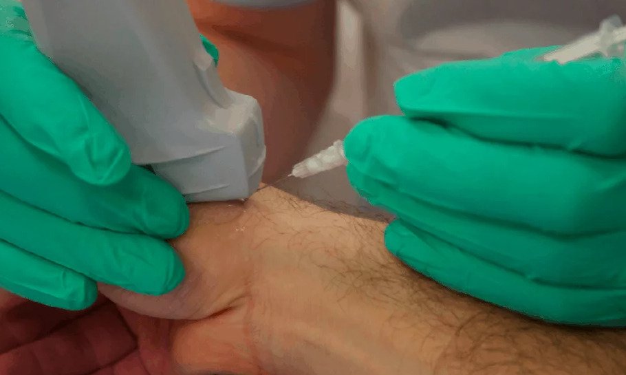
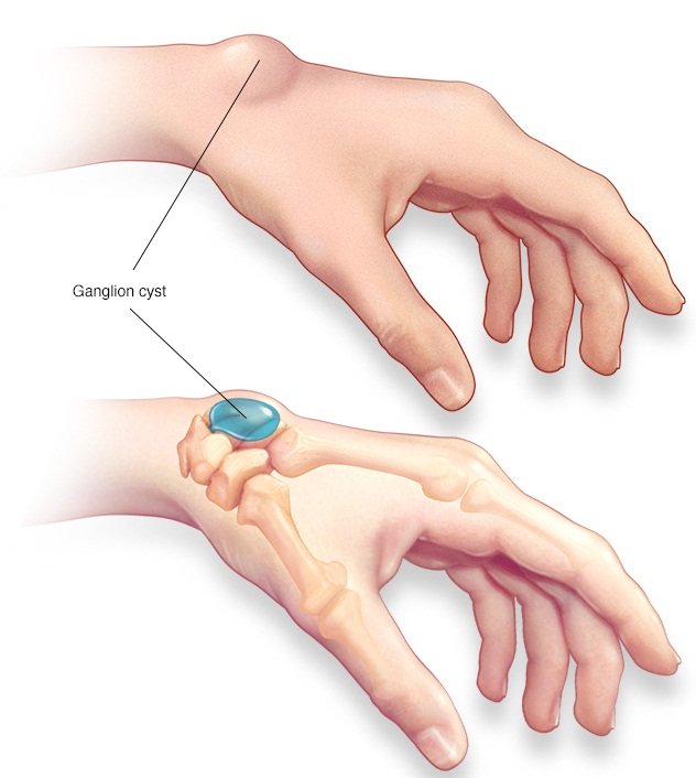
Ultrasound-guided Ganglion cysts assessment

Ultrasound-guided Ganglion cysts assessment

Ganglion cysts are lumps that most commonly develop in the wrist. They’re typically round or oval and are filled with a jelly-like fluid. They are noncancerous tumors that form in the tendons or joints of the wrists or hands. They can also affect the ankles and feet. Ultrasound is often

Read More »
Ultrasound-Guided Medial Collateral Ligament

Ultrasound-Guided Medial Collateral Ligament

The medial collateral ligament (MCL) which also called tibial collateral ligament (TCL), is one of the four major ligaments of the knee. It is on the medial (inner) side of the knee joint in humans and other primates. Its primary function is to resist outward turning forces on the knee. When the outer knee is hit very hard, the MCL, which

Read More »
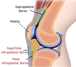
Superficial and Deep Infrapatellar bursa

Superficial and Deep Infrapatellar bursa

Infrapatellar bursitis causes pain and swelling at the front of the knee, just below the knee cap. It develops when there is irritation and inflammation of one of the small fluid-filled sacs in the knee. The infrapatellar bursa is actually made up of two sacs: * Superficial Infrapatellar Bursa: lies between the

Read More »
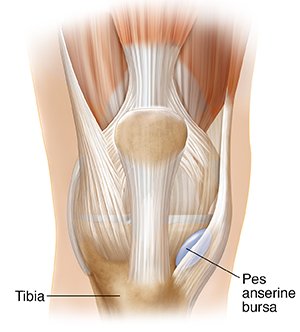
Ultrasound-Guided Anserine bursa

Ultrasound-Guided Anserine bursa

Anserine bursa, also known as intertendinous bursa, is an inflammatory condition of bursa of the conjoined insertion of the sartorius, gracilis and semitendinosus. It is located at the proximal medial aspect of the Knee, two inches below the medial knee joint line between the pes anserinus tendons The anserine bursa is a

Read More »
Suprapatellar recess

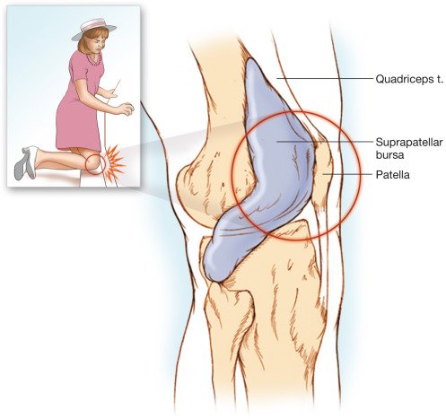
Ultrasound-Guided Suprapatellar recess

Suprapatellar recess also known as the suprapatellar bursa or suprapatellar pouch is one of several bursae of the knee. It is located proximal to the knee joint, between the femoral and suprapatellar fat pads. As with all bursae, its purpose is to reduce friction between moving structures.  Inflammatory joint disorders usually affect the knee

Read More »
Scroll to Top