Ultrasound Guided Facet Nerve Block
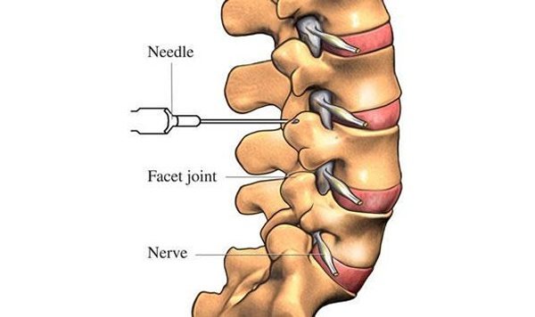
Ultrasound-guided facet nerve block is performed by physicians on patients with chronic back pain by facet arthropathy.
Ultrasound, is a portable, moderately priced imaging modality and is not associated with exposure to radiation.
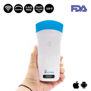
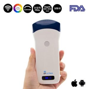
Which ultrasound scanner is used for ultrasound-guided Facet Nerve Block?

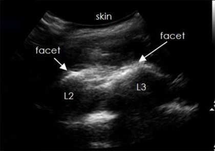
The SIFULTRAS-5.2 not only allows physicians to accurately confirm the surface landmarks of the spinous process and iliac crest line are, But it also obtains longitudinal facet views to identify the different spinal segments.
All needles could be guided successfully to the right segment by ultrasound. Ultrasound guidance is useful in facilitating peripheral and neuraxial blocks and offers direct visualization of the target, adjacent structures, and local anesthetic spread.
The advantages of ultrasound guidance include but are not limited to increased success, decreased rates of complications, faster onset of blocks, and reduced amount of local anesthetics. Ultrasound measurements can even result in suggestions to modify established block techniques.
Ultrasound guided injection provides similar efficacy in the Ultrasound Guided Facet Nerve Block as fluoroscopic guidance but provides benefits of zero radiation and real-time imaging.
Ultrasound guided facet nerve block is usually performed by a radiologist, it may also by performed by a physical medicine and rehabilitation physician (physiatrist).
Reference: Ultrasound-guided Lumbar Facet Nerve Block: A Sonoanatomic Study of a New Methodologic Approach.

[launchpad_feedback]
Although the information we provide is used but doctors, radiologists, medical staff to perform their procedures, clinical applications, the Information contained in this article is for consideration only. We can’t be responsible for misuse of the device nor for the device suitability with each clinical application or procedure mentioned in this article.
Doctors, radiologists or medical staff must have the proper training and skills to perform the procedure with each ultrasound scanner device.


