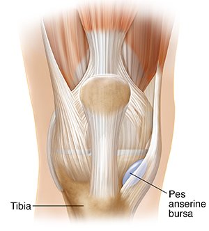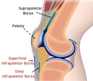
Superficial and Deep Infrapatellar bursa
Infrapatellar bursitis causes pain and swelling at the front of the knee, just below the knee cap. It develops when there is irritation and inflammation of one of the small fluid-filled sacs in the knee. The infrapatellar bursa is actually made up of two sacs: * Superficial Infrapatellar Bursa: lies between the