The Use Of Ultrasound Scanner With Varicose Veins
Varicose veins are veins that are twisted and enlarged. Any superficial vein can become varicose, but the veins in the legs are the most commonly affected. This is due to the fact that standing and walking upright increases the pressure in the lower body’s veins.
Indeed, varicose veins and spider veins — a common, mild variation of varicose veins — are primarily a cosmetic concern for many people. Varicose veins can cause aching pain and discomfort in others. Varicose veins can sometimes lead to more serious problems.
Varicose veins may not cause any pain. Signs a person may have varicose veins include:
· Veins that are dark purple or blue in color
· Veins that appear twisted and bulging; they are often like cords on your legs
Approximately 23% of US adults have varicose veins. If spider telangiectasias and reticular veins are also considered, the prevalence increases to 80% of men and 85% of women. Generally more common in women and older adults, varicose veins affect 22 million women and 11 million men between the ages of 40 to 80 years.
There’s no way to completely prevent varicose veins. But improving the circulation and muscle tone may reduce the risk of developing varicose veins or getting additional ones. The same measures people can take is to treat the discomfort is to see their doctor/phlebologist.
Moreover, medical ultrasound scans are currently the most widely used imaging technique for vascular disease diagnosis. It is frequently used as a varicose veins examination.
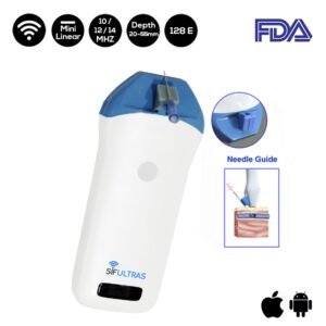
The SIFULTRAS-3.5 Mini Linear Handheld Ultrasound Scanner has numerous advantages, ranging from reducing puncture complications to increasing patient satisfaction.
Doctors can now see blood flow in real-time using an Ultrasound Scanner, which is completely digital and high intensity focused and can provide both qualitative and quantitative data. It is safe to say that without this device, modern phlebology would not exist, and the excellent results we are now seeing with endovenous techniques would not have been possible.
The Mini Linear Handheld Ultrasound Scanner (SIFULTRAS-3.5) is used to image the body’s blood vessels at frequencies of 10MHz / 12MHz / 14MHz and depths of 20mm55mm. It provides detailed information that aids in the confirmation of the diagnosis and pattern of venous disease, as well as the selection of the most appropriate treatment option.
The SIFULTRAS-3.5 has the significant advantage of allowing the phlebologist to monitor the effect of each injection while ensuring the safety of all adjacent structures.
In conclusion, the mini linear ultrasound scanner can detect vein anatomy and flow characteristics. It can aid in the sclerotherapy of veins that are not visible to the naked eye by providing needle guidance and increasing patient safety. It also allows for needle guidance and visualization of deep and superficial venous structures.
References: Varicose veins ,An Introduction to High Intensity Focused Ultrasound , Varicose Veins