
Thoracentesis
A pleural effusion is an abnormal collection of fluid in the pleural space. Removal of this fluid by needle aspiration is called a thoracentesis. The latter can be both diagnostic and therapeutic for the patient.
It is a percutaneous procedure in which a needle or catheter is passed into the pleural space for evacuation of these pleural fluid.
Whereas, therapeutic thoracentesis is performed to palliate dyspnea or impaired ventilation arising from accumulation of pleural fluid, to improve post-drainage chest imaging, to predict success of lung re-expansion in malignant effusions, or to expedite effusion clearance with a single pleural procedure while awaiting the effect of disease-modulating therapies.
Which ultrasound scanner is used for Thoracentesis?
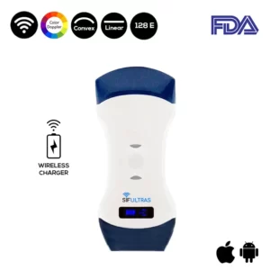
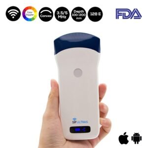
The SIFULTRAS-5.42 is the devise doctors go for since it has both a linear and a curvilenear head.
A curvilinear probe with a frequency of 3.5–5.0 MHz is best suited for performing a thoracentesis. This allows for better visualization of deeper structures and is more than adequate for visualizing more superficial structures adjacent to the chest wall. A higher-frequency probe between 5.0 and 7.0 MHz is effective for visualizing chest wall structures and the parietal pleura.
The sonographer should initially start off deep and survey the chest, the effusion, and surrounding structures. The depth should then be reduced so that the area where the needle will be introduced takes up most of the screen.
The probe can then be moved towards the head and from side to side to locate the largest pocket of fluid between the ribs. Once this is located a mark is made with indelible ink just above the lower rib.
The ultrasound scanner provides improved needle insertion site selection, better characterization of pleural anatomy, and higher rates of procedural success than physical examination alone.
Wireless ultrasound scanner can also precisely identify the location of the fluid so that the chest wall can be marked in preparation for thoracentesis.
In brief, using an ultrasound wand not only detect smaller effusion but most important decrease the very high complication rate associated with it.
The following specialists perform thoracentesis: Pulmonologists, Pediatric pulmonologists, Hospitalists, Critical care medicine doctors, or intensivists, Emergency medicine doctors.


[launchpad_feedback]
Although the information we provide is used but doctors, radiologists, medical staff to perform their procedures, clinical applications, the Information contained in this article is for consideration only. We can’t be responsible for misuse of the device nor for the device suitability with each clinical application or procedure mentioned in this article.
Doctors, radiologists or medical staff must have the proper training and skills to perform the procedure with each ultrasound scanner device.