
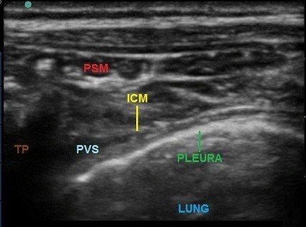
Ultrasound-Guided Thoracic Paravertebral Block
Thoracic paravertebral block (TPVB) is the technique of injecting local anesthetic alongside the thoracic vertebra close to where the spinal nerves emerge from With the use of ultrasound (USG), regional anaesthesia techniques have become safer and with fewer complication rates.
The conventional technique of Thoracic Paravertebral Block involves inserting the needle perpendicular to all planes, contacting the transverse process, and then walking off it with the needle. The commonly-used endpoints for needle insertion include loss-of-resistance to air or saline, advancing a pre-determined distance, or neurostimulation
Which ultrasound scanner is best for Thoracic Paravertebral Block?
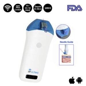
Ultrasound platform with a linear-array transducer oscillating at 14 MHz the SIFULTRAS-3.5 is best for regional ultrasound-guided blocks.
USG helps in avoiding vascular complications and safely placing catheter under anaesthesia. Expertise in USG-guided blocks is essential to ensure success of such procedures.
In conclusion, USG-guided paravertebral block is a valuable armamentarium for post-operative pain management and must be considered following major thoracoabdominal surgeries with unilateral incisions.
Thoracic Paravertebral Block is performed by an Anesthesiologist.

[launchpad_feedback]
Although the information we provide is used but doctors, radiologists, medical staff to perform their procedures, clinical applications, the Information contained in this article is for consideration only. We can’t be responsible for misuse of the device nor for the device suitability with each clinical application or procedure mentioned in this article.
Doctors, radiologists or medical staff must have the proper training and skills to perform the procedure with each ultrasound scanner device.