
Ultrasound for Follicular Growth Monitoring
The study of follicular dynamics and its control has advanced thanks to the use of real-time ultrasonography to monitor ovarian function in mammals. The events that make up follicular development occur in a wave-like pattern. Small (4 to 5 mm) antral follicles grow synchronously to form the waves.
after which one dominant follicle is chosen and grows until it reaches the largest diameter, suppressing the development of the subordinate follicles. The dominant follicle eventually regresses (becomes atretic) in the absence of luteal regression, and a new follicular wave starts. Because the appearance of the subsequent wave is sped up when the dominant follicle is ablated and delayed when its lifespan is extended, the dominant follicle controls the growth of the subordinate follicles.
Bovine oestrous cycles typically have two or three successive waves that appear on days 0 and 10 for cycles with two waves and on days 0, 9, and 16 for cycles with three waves. There are two or three successive dominant follicles during the oestrous cycle, and the last of these ovulates. Pituitary gonadotrophins, ovarian steroids, and non-steroidal elements interact in the complex process of ovarian folliculogenesis. Folliculogenesis is regulated by subtle hormonal changes, and the emergence of a follicular wave is preceded by a slight rise in plasma follicle-stimulating hormone concentration.
Which Ultrasound Scanner Is suitable for follicular growth monitoring?
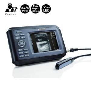
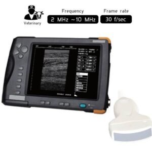
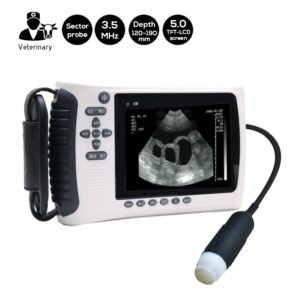
Due to its ready availability, repeatability, and affordability, an ultrasound scanner like the Rectal Veterinary Ultrasound Scanner 3.5 – 5 MHz – SIFULTRAS-4.42 is frequently used as the primary investigation of follicular growth. This is especially true during the pre-stage, which is the diagnosis or assessment before birth.
Additionally, it is employed in patient follow-up because it can clearly illustrate common intraabdominal complications. Another added value, the Waterproof Portable Veterinary Ultrasound Scanner can look at the internal organs. After that, the VET may check the patient for tumors, twists, and other injuries.
Reference: Monitoring follicular development in cattle by real-time ultrasonography: a review
Disclaimer: Although the information we provide is used by different doctors and medical staff to perform their procedures and clinical applications, the information contained in this article is for consideration only. SIFSOF is not responsible neither for the misuse of the device nor for the wrong or random generalizability of the device in all clinical applications or procedures mentioned in our articles. Users must have the proper training and skills to perform the procedure with each ultrasound scanner device.
The products mentioned in this article are only for sale to medical staff (doctors, nurses, certified practitioners, etc.) or to private users assisted by or under the supervision of a medical professional.