Ultrasound Scanner and Pleural Effusion Diagnosis
Pleural effusion, sometimes known as “water on the lungs,” is the accumulation of excess fluid between the layers of the pleura outside the lungs. The pleura are thin membranes that border the lungs and the interior of the chest cavity, lubricating and making breathing easier. The pleura normally contains a little quantity of fluid.
According to the National Cancer Institute, pleural effusions are highly prevalent, with around 100,000 instances identified in the United States each year.
Depending on the cause, the excess fluid may be either protein-poor (transudative) or protein-rich (exudative). These two categories help physicians determine the cause of the pleural effusion.
The most common causes of transudative (watery fluid) pleural effusions include:
- Heart failure
- Pulmonary embolism
- Cirrhosis
- Post open heart surgery
Some patients with pleural effusion have no symptoms, with the condition discovered on a chest x-ray that is performed for another reason. The patient may have unrelated symptoms due to the disease or condition that has caused the effusion. Symptoms of pleural effusion include:
- Chest pain
- Dry, nonproductive cough
- Dyspnea (shortness of breath, or difficult, labored breathing)
- Orthopnea (the inability to breathe easily unless the person is sitting up straight or standing erect)
When it comes to diagnosing pleural effusions, ultrasonography technology is especially helpful when the effusion is tiny or loculated. Ultrasound evaluation enables for the selection of the optimal site for the puncture as well as the assessment of the depth of the nearby organs to avoid organ harm.
In fact, Bedside ultrasound has proved superior to radiography for this condition as it has demonstrated a sensitivity of 93%, compared with 39% for radiography.
Even though studies proved ultrasound scanning efficacy, not all scan devices can provide a clear and accurate diagnosis. That is, a lot depends on the scanning device being used during the examination. It should be professional enough to guarantee a perfectly clear imaging quality that would, later on, help doctors determine in which exact stage the patient is and what’s the best treatment might suit him.
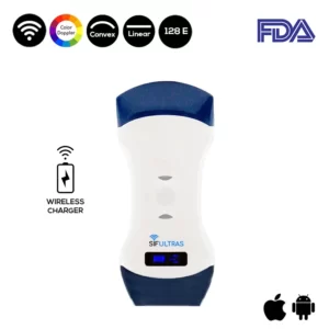
In this context, our technical medical team highly suggests the high-resolution Color Double Head Wireless Ultrasound Scanner SIFULTRAS-5.42 FDA.
This innovative color wireless ultrasound scanner has two heads, thus, making it more practical and more affordable than buying two separate single-headed probes, the two of which are highly needed during this complex examination procedure.
The convex side of the color doppler transducer is used for in-depth examinations of the internal parts of the body like the lungs and so it is convenient for examining Pleural Effusion issue.
In fact, the SIFULTRAS-5.42 ultrasound equipment was created specifically for pulmonologists to produce colored lung images and transfer them to their and their patients’ phones or tablet screens so that both parties are fully aware of the severity of the problem and can discuss the best treatment options in complete transparency.
Plus, the device is IOS and Android compatible. Small and light, easy to carry, and easy to operate. In other words, the SIFULTRAS-5.42 does not compensate for the colored image quality.
To conclude, the SIFULTRAS-5.42 ultrasound equipment should be the top choice for pulmonologists and patients with Pleural Effusion, especially because it is designed to watch vital internal organs such as the lung. As a consequence, patients need not be afraid because they will be shown accurate scan imaging, resulting in a safer and faster assessment. Our medical-technical team strongly recommends the Convex and Linear Color Doppler wireless Double Head Ultrasound Scanner SIFULTRAS-5.42 for patients with Pleural Effusion.
Reference: Pleural Effusion Causes, Signs & Treatment
Disclaimer: Although the information we provide is used by different doctors and medical staff to perform their procedures and clinical applications, the information contained in this article is for consideration only. SIFSOF is not responsible neither for the misuse of the device nor for the wrong or random generalizability of the device in all clinical applications or procedures mentioned in our articles. Users must have the proper training and skills to perform the procedure with each ultrasound scanner device.
The products mentioned in this article are only for sale to medical staff (doctors, nurses, certified practitioners, etc.) or to private users assisted by or under the supervision of a medical professional.

