Ultrasound for vascular access in newborn pigs with Color Doppler
Doppler ultrasonography is used to quantify the direction and velocity of a moving subject. In veterinary medicine, this is most commonly applied to blood flow.
When performing ultrasound tests, the ultrasound wave reflected from the stationary object returns to the transducer at the same frequency at which it was transmitted.
However, if an ultrasound wave is reflected from a moving target, the frequency of the returning echo will be altered by the Doppler effect. This frequency change is known as Doppler Shift and can be detected by an ultrasound machine and displayed as color pixels or in a graphical format.
Doppler ultrasound is most commonly used in echocardiographic examinations. By mapping the velocity and direction of blood flow, areas of abnormal or turbulent blood flow associated with cardiac changes can be visualized.
Besides, the quantification of blood velocity in the heart chambers provides information on changes in blood pressure through the modified Bernoulli equation.
Which Ultrasound Scanner is the best for Vascular access in newborn pigs?
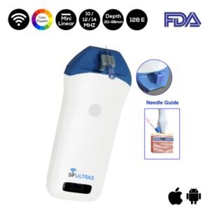
Using a high-frequency ultrasound scanner with a needle guide is more suitable for vascular access in newborn animals and specifically pigs. For this reason, SIFSOF ‘s medical research and development team usually recommend the Color Doppler Mini Linear WiFi Ultrasound Scanner SIFULTRAS-3.51 to our veterinary clients.
The Color Linear Handheld WiFi Ultrasound Scanner 10-12-14 MHz SIFULTRAS-3.51 provides guided injections. It allows the practitioner to visualize the needle in real-time as it enters the body and traverses to the desired location.
Despite good intentions, even in the most experienced hands, blind (injections without imaging) injections are not 100% accurate and in some joints, the accuracy is as low as 30 per cent-40 percent.
With ultrasound guidance, the accuracy of almost every joint injection exceeds 90% and is approaching 100% in many cases.
Indeed, real-time US puncture guidance allows the vet to adjust the vessel’s patency as well as the position of the needle, wire, and catheter in the vessel.
Also, ultrasound can be used to determine the change in the patient’s position and thus the cross-section of the patient’s lumen, depending on the change in the patient’s position.
Moreover, using a Color Doppler ultrasound scanner with a needle guide improves needle placement and injection accuracy, Provides control of the needle insertion path, needle depth, or angle without the risk of damaging adjacent structures, and reduces the time of intravascular access, minimizes the number of unsuccessful catheterization tempts, and reduces the risk of post-puncture complications.
To sum up, Ultrasound guidance is a useful tool to help identify venous and arterial vessels and may contribute to a quick and reproducible venipuncture and vascular access in piglets.
References: Ultrasound Doppler explained for vets,

