Airway Assessment
Airway Assessment is a procedure done to manage patient airway problems (Failure to oxygenate, Failure to ventilate, failure to maintain a patent airway…).
The prediction of difficult mask ventilation (DMV) is a challenging procedure, this is why the Ultrasound Scanner is a helpful tool in the prediction of these difficulties.
Airway-related morbidity, as the result of an inability to anticipate difficult airway, remains the primary concern for anesthesiologist.
Because of high quality of imaging, non-invasiveness and relatively low cost, ultrasonography has been utilized as a valuable adjunct to the clinical assessment of the airway
Which ultrasound scanner is used for airway assessment?
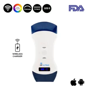
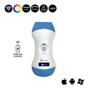
The SIFULTRAS-5.42 is a reliable tool for the assessment of the subglottic airway. The advantages that this Ultrasound offers is that requires minimal training, and doesn’t need patient immobility or sedation.
The newer models are compact and can be rapidly moved with ease from the pre-anesthesia holding area to the operating room. This portability allows point-of-care use of ultrasound US whenever and wherever it is needed. And practicality as it allows anesthesiologists to evaluate complex and varied anatomy.
Since many anesthesia providers had already acquired proficiency in US techniques in US guided vascular access and regional nerve blocks, using US to evaluate the airway could be learned and mastered without too much difficulty. Ultrasound of the upper airway may prove to become a useful adjunct to conventional clinical assessment tools, as it has been successful in visualizing the relevant anatomy and critical structures of the airway
This device allows the doctor to estimate the appropriate tracheostomy the size and length and avoid anterior neck structures as well as posterior tracheal wall injuries.
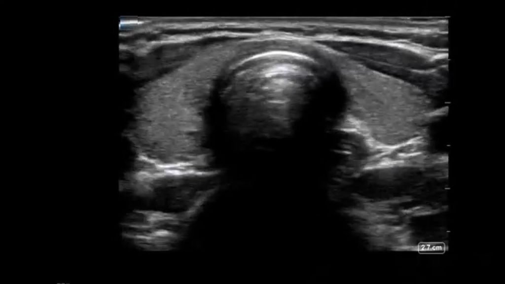
In the preoperative holding area sonographic assessment is done with the help of a high-frequency linear probe. By placing the probe in the submandibular area in the midline. Without changing the position of the probe, the linear array of the US probe was rotated in the transverse planes from cephalad to caudal, until simultaneous visualization of the epiglottis and posterior part of vocal folds with arytenoids observed on the screen. Thereafter, following measurements are obtained with the oblique-transverse US view of the airway .
Similarly, curved low-frequency (5 MHz) transducer is used to visualize the tongue and shadows of the hyoid bone and mandible with the patient in the supine position. The hyomental distances is measured from the upper border of the hyoid bone to the lower border of the mentum in the neutral and extended head positions, respectively.
Using ultrasound to help assess the difficult airway constitutes just yet another valuable application of this versatile technology.
Reference: Ultrasound-Assisted Evaluation of the Airway in Clinical Anesthesia Practice: Past, Present and Future.
[launchpad_feedback]
Although the information we provide is used but doctors, radiologists, medical staff to perform their procedures, clinical applications, the Information contained in this article is for consideration only. We can’t be responsible for misuse of the device nor for the device suitability with each clinical application or procedure mentioned in this article.
Doctors, radiologists or medical staff must have the proper training and skills to perform the procedure with each ultrasound scanner device.


