Deep Vein Thrombosis preview

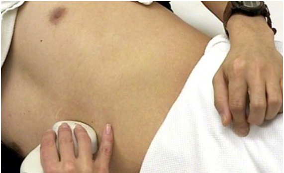
DVT : Deep Venous Thrombosis

A deep venous thrombosis DVT ultrasound is used to image or see the clot. This examination purpose is to evaluate clot in the femoral and popliteal regions. Which ultrasound scanner is used for Deep Venous Thrombosis diagnosis? Using the SIFULTRAS-5.31 while imaging the deep veins of the leg, the physician

Read More »
Appendicitis Abnormal Appendix

Abnormal Appendix : Appendicitis

Appendicitis : Abnormal Appendix, is an inflammation of the vermiform appendix, a closed tube of tissue attached to the large intestine in the lower right abdomen. Inflammation may occur when the appendix becomes infected or blocked with stool, by obstruction of the appendiceal lumen, with the resultant build-up of fluid,

Read More »
Ultrasound guided lumbar central neuraxial block

CNB : Central Neuraxial Block

The practice of central neuraxial block (CNB) has traditionally relied on the palpation of bony anatomical landmarks, namely the iliac crests and spinous processes, together with tactile feedback during needle insertion. However, these landmarks may be difficult to identify accurately—a problem exacerbated by altered patient anatomy, including obesity, age-related changes,

Read More »
Transthoracic Echocardiogram TTE

TTE : Transthoracic Echocardiogram

A transthoracic echo TTE is a clinical procedure that’s done to evaluate the structure and function of the heart. It is the most common noninvasive type of echocardiogram, which uses high frequency soundwaves (ultrasound) to create a moving picture of your heart through the chest wall. Which ultrasound scanner is

Read More »
FOCUS Focused Cardiac Ultrasound

Focused Cardiac Ultrasound : FOCUS

Patients with undifferentiated shock, hypotension, chest pain, or dyspnea are ideal candidates for focused cardiac ultrasound (FOCUS). In addition, any patient in whom the diagnosis of pericardial effusion or tamponade, pulmonary embolism, or left ventricular dysfunction is suspected would benefit from FOCUS. Which ultrasound scanner do doctors choose for Focused

Read More »
Ultrasound of the abdomen

Abdominal Pelvic Scan

[vc_row][vc_column][vc_column_text] An abdominal pelvic scan, is a rather frequent diagnostic procedure, that gynecologists perform on a day to day basis. Using the right ultrasound scanner to assess organs and structures within the female pelvis. A pelvic ultrasound allows quick visualization of the female pelvic organs and structures including the uterus,

Read More »
FAST - Focused Assessment with Sonography in Trauma - SIFSOF

FAST: Focused Assessment with Sonography in Trauma

The Focused Assessment with Sonography in Trauma (FAST) is a point-of-care ultrasound examination performed at the time of presentation of a trauma patient. It is an ultrasound protocol developed to identify intraperitoneal free fluid (assumed to be hemoperitoneum in the context of trauma) allowing for an immediate transfer to theater,

Read More »
A pleural effusion is an abnormal collection of fluid in the pleural space. Removal of this fluid by needle aspiration is called a thoracentesis. The latter can be both diagnostic and therapeutic for the patient.

Thoracentesis

A pleural effusion is an abnormal collection of fluid in the pleural space. Removal of this fluid by needle aspiration is called a thoracentesis. The latter can be both diagnostic and therapeutic for the patient. It is a percutaneous procedure in which a needle or catheter is passed into the

Read More »
GUS Gastric Ultrasound Scanner

GUS : Gastric Ultrasound Scanner

Patients with comorbidities that delay gastric emptying, such as diabetic gastroparesis, neuromuscular disorders, morbid obesity, and advanced hepatic or renal disease, may potentially benefit from additional assessment via gastric ultrasound scanner (GUS) before an elective procedure. The main objective of point-of-care (POC) gastric ultrasound is to help clinicians assess gastric

Read More »
Scroll to Top