CNB : Central Neuraxial Block
The practice of central neuraxial block (CNB) has traditionally relied on the palpation of bony anatomical landmarks, namely the iliac crests and spinous processes, together with tactile feedback during needle insertion.
However, these landmarks may be difficult to identify accurately—a problem exacerbated by altered patient anatomy, including obesity, age-related changes, and previous spinal surgery.
Nonetheless, detailed knowledge of lumbar spinal anatomy and sonoanatomy is essential for interpretation of neuraxial ultrasound images.
Which ultrasound scanner is used for central neuraxial block?
The most suitable wireless ultrasound probe used for CNB is a curved probe of low frequency the SIFULTRAS-5.2 as it allows deeper penetration at the expense of image resolution.
In brief, Ultrasound-assisted CNB is an advanced technique for use in patients with difficult spinal anatomy. That is why, performing a pre-procedural scan helps to identify relevant landmarks and thus guide subsequent needle insertion.
Compared to palpation of surface anatomical landmarks, pre-puncture ultrasound scan can identify the vertebral levels more accurately.
The use for CNB allows for any anatomical variances to be seen and their possible complicating effects adjusted for. In addition, it has had a significant reduction in both skin punctures and needle redirection attempts with the use of ultrasound.
The use of the portable ultrasound as pre-procedural scan by the anesthesiologist also improves the technical efficiency of CNB by facilitating precise identification of underlying anatomical structures.
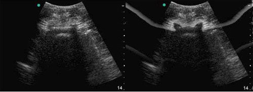
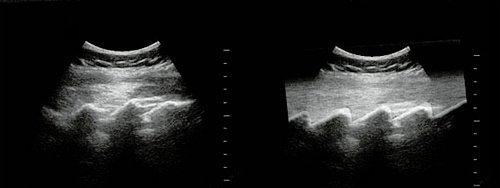
Pre-puncture ultrasound guidance is easy to perform and hence gaining popularity, which involves first performing a “scout ultrasound scan” to assess the target vertebral level and ligamentum flavum-dura complex, and adjacent anatomy.
[launchpad_feedback]
Disclaimer: Although the information we provide is used by different doctors and medical staff to perform their procedures and clinical applications, the information contained in this article is for consideration only. SIFSOF is not responsible neither for the misuse of the device nor for the wrong or random generalizability of the device in all clinical applications or procedures mentioned in our articles. Users must have the proper training and skills to perform the procedure with each ultrasound scanner device.
The products mentioned in this article are only for sale to medical staff (doctors, nurses, certified practitioners, etc.) or to private users assisted by or under the supervision of a medical professional.

