Diagnostic Accuracy of Ultrasound for Assessment of Arthropathic hemophilia
The hemophilia is a combination of the greek word “blood” and “love” a way of saying that people with hemophilia “love to bleed” or rather or rather that it’s hard to stop bleeding. This is because the process called hemostasis, literally meaning to stop the flow of blood is impaired. Normally after a cut or damage to the endothelium or lining of blood vessels walls there’s an immediate vasoconstriction or narrowing of the blood vessels which limits the amount of blood flow. After that some platelets adhere to the damaged vessel wall, and become activated and then recruit additional platelets to form a plug. The formation of this platelet plug is called primary hemostasis. After that the coagulation cascade is activated. First Off the blood has a set of clotting factors. Most of which are proteins synthesized by the liver and usually these are inactive and just floating around in the blood. The coagulation cascade starts when one of these proteins gets proteolytically cleaved. This active protein then proteolytically cleaves and activates the next clotting factor and so on. This cascade has a great degree of amplification and takes only a few minutes from injury to clot formation. The final step is activation of the protein fibrinogen (factor1) to fibrin. Which deposits and polymerizes to form a mesh around the platelets. So these steps leading up to fibrin reinforcement of the platelet plug make up the process called secondary hemostasis and result in hard clot at the site of the injury.
In most cases of hemophilia there is a decrease in the amount or function of one or more of the clotting factors which makes secondary hemostasis less effective and allows more blessing to happen. Now that coagulation cascade can start in either two ways. First way is called the extrinsic pathway, which starts when tissue factor gets exposed by the injury of the endothelium. Tissue factor turns inactive factor 7 into active factor 7A (A for active), and then tissue facto goes on to bind with the newly formed factor 7A to form a complex that turns factor 10 into active factor 10A. Factor 10A with factor 5A as a cofactor turns factor 2 which is also (which is also called prothrombin) into factor 2A which is also called thrombin. Thrombin then turns factor 1 or fibrinogen which is soluble into 1A or fibrin which is insoluble and precipitates out of the blood at the site of injury. Thrombin also turns factor 13 into factor 13A which crosslinks the fibrin to form a stable clot. The second way is called the intrinsic pathway and starts with platelets near the blood vessel injury activates factor 12 into factor 12A which then activates factor 11 into factor 11A which then activates factor 9 into factor 9A. Factor 9A along with factor 8A work together to activate factor 10 to factor 10A and from that point it follows the same fate as before. So the extrinsic and intrinsic pathways basically converge on a single final path called the common pathway. This is a somewhat simplified version of the coagulation cascade but it has all the key parts needed to understand hemophilia. An insufficient or decreased activity of any coagulation factor can cause hemophilia except factor 12 deficiency which is asymptomatic.
Hemophilia usually refers to inherited deficiencies, either quantitative or qualitative. By far the most common of these are factor 8 which gives rise to factor 8A and is stabilized by another factor called von wilebrand factor. This deficiency is called hemophilia A or classic hemophilia. Another common deficiency is deficiency in factor 9 called hemophilia B which used to be called Christmas disease named after the first person who had it not the holiday.
Hemophilia patients require lifelong clotting factor replacement therapy to mitigate spontaneous joint bleeding and other life threatening bleeding. However, clotting factor replacement therapy is costly and imposes a high financial burden on individuals, healthcare systems and society in general. Joint bleeding represents the most commonly reported type of hemorrhage in patients affected by hemophilia. Although the widespread use of prophylaxis has been able to significantly reduce the onset of arthropathy, it has been shown that a non-negligible percentage of patients develop degenerative changes in their joints despite this type of treatment. Thus, periodic monitoring of the joint status in hemophilia patients has been recommended to identify early arthropathic changes and prevent the development or progression of hemophilic arthropathy. Ultrasound (US) has proven able to detect and quantify the most relevant biomarkers of disease activity (i.e., joint effusion and synovial hypertrophy) and degenerative damages (i.e., osteo-chondral changes) by means of scoring scales of increasing disease severity. Therefore, timely objective detection of acute or persistent joint bleeding in hemophilia patients has become increasingly important.
Magnetic resonance imaging (MRI) is considered the “gold standard” to detect various abnormalities in hemophilic arthropathy. However, in the past few years, musculoskeletal ultrasound (MSKUS) has emerged as a point-of-care (POC) imaging tool to assess the extent of arthropathic changes, thus opening new avenues for the management of hemophilic arthropathy and also rapid joint bleed detection. Recent advances in technology, accessibility, and training have made POC MSKUS an attractive alternative to MRI in instances where imaging is desired. MSKUS is faster, more economical, and without the need of sedation for claustrophobic subjects or children. In addition, MSKUS does not require intravenous contrast to distinguish synovial proliferation from fluid and can also be used to assess synovial vascularity.
MSKUS appears very adept in detecting joint effusions based on the ability of dynamic maneuvers during scanning. For hemophilia, this feature appears particularly valuable for the detection and management of hemarthrosis, where precise diagnosis of presence or absence of (bloody) effusions can complement patient or physician perception, thereby optimizing targeted treatment options. It allows visualization of shifting fluid in communicating spaces as well as sonopalpation.
Sonopalpation assesses compressibility and displacement of echogenic intra-articular material. Effusions can be separated into simple versus complex. Complex fluid accumulations are characterized by mixed echogenicity and displaceable speckles, indicating the presence of particulate matter such as proteins or blood products, while simple effusions appear anechoic with clear and serous fluid upon aspiration. Thus, MSKUS not only documents the presence of an effusion, but also distinguishes between bloody versus non-bloody effusions based on echogenicity (echogenic versus anechoic) and presence of displaceable echogenic reflectors.
In the context of hemophilia, complex effusions with echogenic reflectors can be assumed to represent blood products based on previous documentation of the great accuracy of this approach as documented by joint aspiration. MSKUS algorithms to detect hemarthrosis are therefore well defined, and can be performed quickly as part of the daily clinic routine, thereby fulfilling POC criteria. Moreover, MSKUS enables guided aspiration and fluid analysis as clinically indicated.
In this context, it is noteworthy that radiological MRI criteria to assess blood contents in the joint are less well defined, and mainly derived from previous neurological studies. A preliminary study 30 years ago suggested that MRI may not have the same usefulness for distinguishing between bloody and non-bloody effusions in joints. However, formal studies employing modern imaging technology are lacking, and clinical imaging interpretation algorithms more commonly use inference rather than evidence. Moreover, in daily clinical practice, joint effusions on MRI automatically may be deemed as bloody if arising in the context of hemophilia.
MSKUS has proven to be extremely sensitive in detecting low concentrations of intra-articular blood and in discriminating between bloody and non-bloody fluid, whereas conventional MRI is not. These observations demonstrate the advantages of MSKUS over MRI in detecting intra-articular blood, and show that MSKUS is ideal for rapid bleed detection in the clinic.
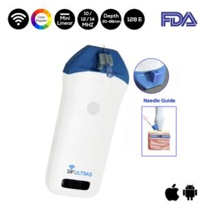
For this type of diagnoses we highly recommend the Color 5-10 MHz Wireless Linear Ultrasound Scanner 128 Elements SIFULTRAS-5.38. This ultrasound Resolution visualizes delicate tissue structure in shallower regions. It’s image clarity reduces the noise in blood vessels at a frequency range of 5-10 MHz frequency and 40-120mm depth. , the SIFULTRAS-5.38 does not only serve hemarthrosis but orthopedy in general. The Color Linear ultrasound scanner provides qualitative and quantitative for musculoskeletal diagnosis. For example: Tendon tears, or tendinitis of the rotator cuff in the shoulder, Achilles tendon in the ankle and other tendons throughout the body, muscle tears, masses or fluid collections., ligament sprains or tears.
Using the SIFULTRAS-5.38 the physician can detect; inflammation or fluid (effusions) within the bursae and joints, early changes of rheumatoid arthritis, nerve entrapments such as carpal tunnel syndrome, benign and malignant soft tissue tumors, ganglion cysts, hernias., foreign bodies in the soft tissues (such as splinters or glass), dislocations of the hip in infants, fluid in a painful hip joint in children, neck muscle abnormalities in infants with torticollis (neck twisting), soft tissue masses (lumps/bumps) in children.
This procedure is performed by a qualified orthopedist trained in ultrasound imaging.*

