
Ultrasound-guided Endovenous Laser Ablation (EVLA)
Veins of the lower limb are divided into 2 types: Deep and superficial veins. Due to various factors, the veins can become enlarged and twisted near the surface of the skin.
In the majority of cases, this condition is hereditary. However, other factors can lead to a higher risk of having varicose veins, such as: age, obesity, jobs that involve a long period of standing or sitting, sedentary lifestyle, pregnancy, thrombosis or thrombophlebitis.
Varicose veins are defined as veins visible under the skin, with a diameter greater than 3-4 mm, that appears swollen, palpable and tortuous. Varicose veins are most commonly found in the lower limbs (i.e. legs and ankles).
More than 80% of varicose veins visible on the legs are caused by superficial venous insufficiency of the great saphenous vein, while the sapheno-femoral junction is the main point of reflux in most patients with superficial venous insufficiency.
In order to close off a varicose or an enlarged swollen vein, an Endovenous Laser Ablation (EVLA) should be performed.
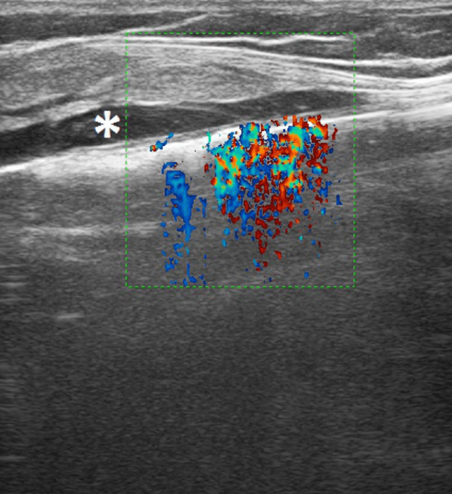
EVLA is a procedure where the doctor uses laser radiation to heat the vein walls in order to create a typical signal with the Color-Doppler imaging.
Using ultrasound scanning is one of the most important non-invasive investigations used in the assessment of vascular problems, including varicose veins. It’s used in evaluating the properties and the state of the patient’s veins.
Contrast-enhanced ultrasound imaging allows the assessment of micro-circulation and can be used for detecting the veins and the points where they intersect.
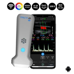
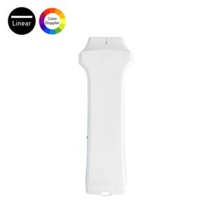
SIFULTRAS-5.17 and SIFULTRAS-5.34 are 2 instances of ultrasound scanners with portable linear color Doppler probes that can be used in Endovenous Laser Ablation (EVLA).
SIFULTRAS-5.17 has a frequency of 4-12 MHz, while SIFULTRAS-5.34 has a frequency of 7.5-10 MHz. Both probes have a scan depth of 20-100 mm.
These 2 ultrasound scanners by SIFSOF give a clear and accurate scan results for superficial veins, making them very suitable for vascular surgeons while performing EVLA.
Ultrasound-guided EVLA of lower limb varicose veins using SIFSOF ultrasound scanners is an effective way that leads to a satisfying clinical outcome. It is a minimally invasive and safe interventional procedure, related to a low risk of complications.
Ultrasound EVLA does not require general anesthesia, so it can be performed in an ambulatory setting, increasing the patient’s comfort and resulting in a faster and cheaper treatment compared to the traditional surgery.
EVLA is almost painless and it is a well-accepted treatment option in patients with superficial venous insufficiency. It’s performed by phlebologists.


References: Ultrasound Guided (US) Endovenous Laser Ablation (EVLA), Duplex Ultrasound Investigation of the Veins of the Lower Limbs, Endovenous Ablation.
[launchpad_feedback]
Disclaimer: Although the information we provide is used by different doctors and medical staff to perform their procedures and clinical applications, the information contained in this article is for consideration only. SIFSOF is not responsible neither for the misuse of the device nor for the wrong or random generalizability of the device in all clinical applications or procedures mentioned in our articles. Users must have the proper training and skills to perform the procedure with each ultrasound scanner device.
The products mentioned in this article are only for sale to medical staff (doctors, nurses, certified practitioners, etc.) or to private users assisted by or under the supervision of a medical professional.