High-frequency Ultrasound in Clinical Dermatology
Today, high-frequency ultrasound (HFUS), (10MHz and above), provides for high-resolution imaging of the skin from the stratum corneum to the deep fascia. It’s a noninvasive and easy-to-interpret screening tool that allows physicians to assess skin conditions in real time, leading to enhanced diagnostic, management, and surgical capabilities. Dermatology is a rare specialty of medicine where the organ of interest can be readily examined and biopsied. Consequently, histology has become the gold standard for many dermatologic diagnoses. It has also allowed for clinicopathologic correlation to guide therapeutics and improve prognostics. In the last few decades, a number of imaging modalities have been introduced to enhance clinical exams and eliminate the need for histology. One example is total body digital photography (TBDP), dermoscopy, reflectance confocal microscopy (RCM), optical coherence tomography (OCT), and ultrasound.
As ultrasound technology has advanced, so has its utility. Compared to clinical examination alone, ultrasound has improved the accuracy of the referring diagnosis. The usage of ultrasound in dermatology is continuously evolving and increasingly studied.
Given the diverse range of probe frequencies and the variation of dermal thickness in different regions of the body, specific frequencies should be considered for optimal visualization of lesions in various anatomic locations.
The recent advancement in point-of-care High-frequency ultrasound imaging has opened a new venue for the diagnosis of skin conditions such as skin inflammatory diseases like Sclerosing disorders, psoriasis, Hidradenitis suppurativa, vascular lesions, and cosmetic applications. Standardized markers like increased and decreased echogenicity and specific anatomical differences to each skin disorder have been established.
Color Doppler, yet another ultrasound imaging modality has shown promise for certain skin conditions. Alegre-Sanchez et al. found that spider nevi demonstrated pulsatile flow on dermoscopy and ultrasound. This finding was subsequently supported by the presence of high-flow arterial-type waveform as seen on Doppler-ultrasound, suggesting that spider nevi are more similar to arteriovenous malformations, or high-flow vascular lesions.
In a nutshell, while ultrasound has been used in dermatology for more than 40 years, its usage is continuously evolving as technology advances. , the DERMUS group suggests utilizing ultrasound at least 300 times per year to achieve minimum competency. Similarly, it is strongly advised that the dermatologist functions as the sonographer because of the years of training in dermatologic pathologies and the ability to correlate histology with the clinical presentation of cutaneous lesions. As frequency advancements in ultrasonography continue, the broad applications of this imaging modality will continue to grow. Ultrasound is a fast, safe, and readily available tool that can aid in diagnosing, monitoring, and treating dermatologic conditions by providing more objective assessment measures.
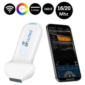
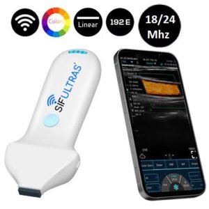
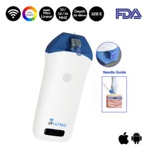
When it comes to clinical dermatology, high-frequency ultrasound imaging, and plastic surgery, we highly recommend the 18/24Mhz Facial Linear Ultrasound Scanner 192 Elements SIFULTRAS-3.35 or Aesthetic Linear Ultrasound machine 16/20Mhz 192 Elements SIFULTRAS-3.36. Tests have shown that the impact of treatment such as laser skin rejuvenation can be detected earlier with ultrasound, and the technology can validate new devices and treatments. The Color Doppler Mini Linear WiFi Ultrasound Scanner SIFULTRAS-3.51 (10 MHZ / 12MHz / 14 MHz) enables clinicians and patients to see the impact of treatments often before visual signs are apparent. Furthermore, this wireless scanner can be a powerful marketing tool for any aesthetics center, as it’s able to show the impact of treatments in a new way and retain records for future visits. Since it is easy to operate it is also suitable for personal use. It is highly useful in plastic surgeries as it provides more accurate needle placement for professional procedures thanks to the needle guide accessory.
The covered imaging and diagnostic procedures are performed by a qualified dermatologist who is trained in ultrasound imaging modalities*
Reference: High-frequency ultrasound in clinical dermatology: a review
Assessment of an ultrasonic dermal scanner for skin thickness measurements