Lymphaticovenular anastomosis LVA
The planning of lymphaticovenular anastomosis LVA is a difficult task for surgeons as it is not easy to observe the lymphatic collectors by the indocyanine green lymphography.
Which ultrasound scanner is best for Lymphaticovenular anastomosis LVA?
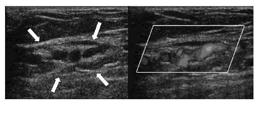
Color Doppler Ultrasound allows you to see both lymphatic collectors and venules. When indocyanine green lymphography cannot locate any collector, color Doppler ultrasound will help in its visualization.
Even with the venule, Color Doppler Ultrasound is better than other tools because it gives the exact location of the venule, to set its standard so it can match with the collector and to preoperatively evaluate the absence of blood reflux within the selected venule for the presence of a valve.
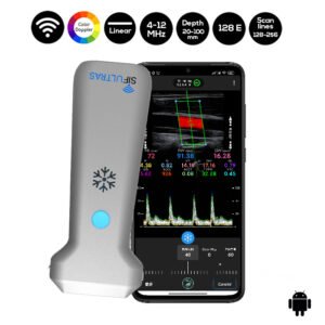
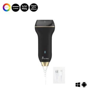
To do this examination the practitioner needs a linear Doppler Ultrasound with a depth of 2 to 3 cm. Doppler ultrasound scanner SIFULTRAS-5.34 linear probe with 20 to 100mm scanning depth.
Accordingly it is very useful in which the lymphaticovenular anastomosis surgeon can plan skin slit more efficiently and provides decrease in operative exploration time.
This procedure is performed by Lymphologists, lymphaticovenular anastomosis surgeons…

[launchpad_feedback]
Although the information we provide is used but doctors, radiologists, medical staff to perform their procedures, clinical applications, the Information contained in this article is for consideration only. We can’t be responsible for misuse of the device nor for the device suitability with each clinical application or procedure mentioned in this article.
Doctors, radiologists or medical staff must have the proper training and skills to perform the procedure with each ultrasound scanner device.


