
M-Mode Ultrasound Imaging for Identifying a Pneumothorax
A pneumothorax is the collapse of a lung. A pneumothorax occurs when air enters the space between the lung and the chest wall. This air presses on the lung’s outer surface, forcing it to collapse. It might be a complete or partial collapse of the lungs.
A penetrating or severe chest injury, certain medical procedures, or lung sickness can all produce a pneumothorax.
It might also happen for no apparent cause. Typical symptoms include severe chest discomfort and loss of breath. A collapsed lung can be life-threatening in some cases.
The use of ultrasound imaging has a significant benefit in point-of-care settings, and it appears to have a high sensitivity for detecting a pneumothorax.
In fact, the presence of sonographic imaging is a sensitive indicator for detecting a pneumothorax. M-mode ultrasonography can be a helpful adjuvant in detecting the disease.
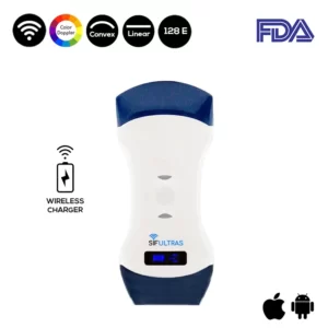
our technical medical team strongly suggests the Color Double Head Wireless Ultrasound Scanner SIFULTRAS-5.42 FDA, which may be utilized for many applications at once, most notably in pulmonology.
Because this one-of-a-kind color wireless ultrasound scanner has two heads, it is more convenient and less expensive than buying two separate single-headed probes.
The color doppler transducer’s convex side is used for comprehensive examinations of internal organs such as the lungs, and its display modes: M and B with a gray scale of 256 levels make it simple to assess the pneumothorax condition.
Actually, the use of color Doppler sonography as an adjuvant to B- and M-mode imaging for pneumothorax identification has been documented in a few studies and case reports.
Indeed, the SIFULTRAS-5.42 Ultrasound Probe is specifically designed for pulmonologists to generate colored lung images and transfer them to their and their patients’ phones or tablet screens, so that both parties are fully aware of the gravity of the situation and can discuss the best treatment option in complete transparency.
Furthermore, the gadget is compatible with both iOS and Android. Small and light, convenient to transport and use. In other words, the Wireless Ultrasound Scanner SIFULTRAS-5.42 does not adjust for the quality of colorful images.
To recap, the use of B- and M-mode sonography for pneumothorax detection has been extensively reported on and investigated. It is being used by sonographers, emergency doctors, trauma surgeons, radiologists, and critical care specialists all around the world. Lung sonography can be performed immediately at the patient’s bedside or in the emergency room. It is more sensitive, specific, and accurate than standard chest radiography.
References: collapsed lung,
Disclaimer: Although the information we provide is used by different doctors and medical staff to perform their procedures and clinical applications, the information contained in this article is for consideration only. SIFSOF is not responsible neither for the misuse of the device nor for the wrong or random generalizability of the device in all clinical applications or procedures mentioned in our articles. Users must have the proper training and skills to perform the procedure with each ultrasound scanner device.
The products mentioned in this article are only for sale to medical staff (doctors, nurses, certified practitioners, etc.) or to private users assisted by or under the supervision of a medical professional.