Musculoskeletal Ultrasound (MSK)
Ultrasound images of the musculoskeletal (MSK) system provide pictures of muscles, tendons, ligaments, joints, nerves and soft tissues throughout the body. The diagnosis of tissues is a specific process. MSK ultrasound requires top-class equipment with the highest quality transducers.
Which Ultrasound scanner do doctors use for Musculoskeletal?
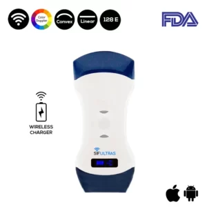
Two wireless ultrasound scanners are suitable for MSK applications: the SIFULTRAS-5.31 and SIFULTRAS-5.34. These two portable ultrasound machines gain full software options to improve image quality, resolution, contrast and artifact removal.
The SIFULTRAS-5.31 provides black and white images for different MSK application scenarios. Whereas, the SIFULTRAS-5.34 offers Doppler options: color, power and tissue (microcirculation) imaging. Besides that, The SIFULTRAS-5.42 has a Convex side which can be used for imaging obese patients or examining deep structures.
Musculoskeletal ultrasound (MSK) involves not only the imaging of soft tissues throughout their available range, but also the visualization of elements structurally or functionally connected with them.
e.g. the examination of muscle-tendon units should include the tendons at the muscle belly level and the naked (bare) part, their entheses, as well as all peritendinous elements, such as the peritendineum, sheaths, retinaculum, bursa, fascia, subcutaneous tissue, fat folds or bone outlines, main vessels and regional nerves.
Ultrasound images are typically used to help diagnose: tendon tears or tendinitis of the rotator cuff in the shoulder, Achilles tendon in the ankle and many other tendons throughout the body. muscle tears, masses or fluid collections. ligament sprains or tears. inflammation or fluid (effusions) within the bursae and joints.
Early changes of rheumatoid arthritis. nerve entrapments such as carpal tunnel syndrome. benign and malignant soft tissue tumors. ganglion cysts. hernias. foreign bodies in the soft tissues (such as splinters or glass). dislocations of the hip in infants. fluid in a painful hip joint in children. neck muscle abnormalities in infants with torticollis (neck twisting). soft tissue masses (lumps/bumps) in children.
Musculoskeletal ultrasound is done by a radiologist, physical therapist, sports doctor, MSK doctor.

[launchpad_feedback]
Although the information we provide is used but doctors, radiologists, medical staff to perform their procedures, clinical applications, the Information contained in this article is for consideration only. We can’t be responsible for misuse of the device nor for the device suitability with each clinical application or procedure mentioned in this article.
Doctors, radiologists or medical staff must have the proper training and skills to perform the procedure with each ultrasound scanner device.


