Nonalcoholic Fatty liver Disease Bedside Ultrasound Diagnosis
The thyroid is a butterfly-shaped gland that sits low on the front of the neck. The thyroid lies below Adam’s apple, along the front of the windpipe. It has two side lobes, connected by a bridge (isthmus) in the middle. The thyroid secretes several hormones, collectively called thyroid hormones. The main hormone is thyroxine, also called T4. Thyroid hormones act throughout the body, influencing metabolism, growth and development, and body temperature. During infancy and childhood, adequate thyroid hormone is crucial for brain development. Clinical trials have established a link between hypothyroidism and nonalcaholic fatty liver disease.
Steatosis, or fatty liver, is a common result of moderate to severe insult to Hepatocytes which are major parenchymal cells in the liver which play a pivotal roles in metabolism, detoxification, and protein synthesis. Hepatocytes also activate innate immunity against invading microorganisms by secreting innate immunity proteins.
An underactive thyroid causes the metabolism to slow down and leads to an increase in thyroid stimulating hormone (TSH) levels. This also leads to an accumulation of fat in the body, which increases the risk of developing non-alcoholic fatty liver disease (NAFLD).
In the last 20 years, nonalcaholic fatty liver disease has become one of the most common liver diseases in the world, encompassing almost 25% of the world’s population including children.
Hepatic steatosis is diagnosed by ultrasound after exclusion of infectious and metabolic disorders. While ultrasound diagnosis and fasting serum samples are taken for determination of thyroid function (TSH, FT4, and FT3), along with alanine aminotransferase (ALT), lipid profile, glucose, insulin, and insulin resistance.
Ultrasound is a non-invasive, widely available, and accurate tool in the detection of Non-alcoholic fatty liver disease (NAFLD). Ultrasound should be used as the first-line diagnostic test in patients with abnormal liver enzymes when other causes are excluded. Clinical risk factors, when used with ultrasound findings, have high accuracy in identifying NAFLD patients. There is an an algorithm for chronic abnormal liver enzymes that illustrates the use of ultrasound in reducing the need for liver biopsy in the diagnosis of NAFLD. Although Clinicians should be aware of the known limitations of ultrasound, including the inability to grade or stage fibrosis in NAFLD patients.
For the above mentioned ultrasound applications we recommend the following two ultrasound transducers:
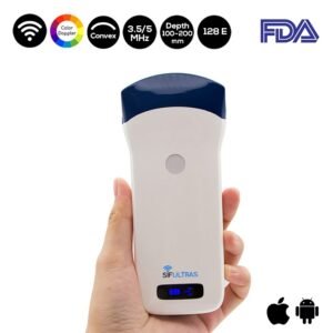
- Color Doppler Wireless Convex Ultrasound Scanner 3.5-5MHz, SIFULTRAS-5.21. This device is best suitable for detection of non alcaholic fatty liver disease. It allows you to visualize liver function and make diagnoses quickly and confidently.
- Linear Wireless Ultrasound Scanner SIFULTRAS-5.34 – equipped with a high resolution 7.5 MHz transducer this device is recommended for measuring thyroid size and morphology with the subjects sitting and their necks slightly extended. Thyroid volume is defined as enlarged according to the reference values for age, gender, and body surface area.
This procedure is performed by an elligible Endocrinologist
Reference: Hypothyroidism and Nonalcoholic Fatty Liver Disease: Pathophysiological Associations and Therapeutic Implications
Hepatic Steatosis and Thyroid Function Tests in Overweight and Obese Children


