Ultrasound-Guided Foreign Body Retrieval
Historically, removal of intravascular foreign bodies ( FBs) in veterinary patients had been performed via surgical extraction.
The majority of intravascular foreign bodies are caused by the dislodgement of intravascular catheters or other equipment inserted into the vasculature during the conduct of a specific intervention.
The complication rate linked with intravascular FB retrieval is minimal, with the most common complications being a failure to retrieve and transient arrhythmias.
The techniques most commonly employed to grasp an intravascular foreign body include the snare loop technique, the use of helical baskets and the use of intravascular forceps/graspers.
Indeed, a study has shown that The surgical removal of FBs can be intrusive, expensive, and technically difficult. Yet, Ultrasound has become a standard in the identification of Foreign Bodies and may be utilized to guide the extraction of Foreign Bodies using a minimally invasive method.
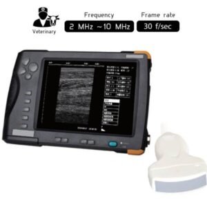
For Example, The Portable Veterinary Ultrasound Scanner 2-10MHz Waterproof SIFULTRAS-4.2 is highly recommended to be used by vets, due to its Working frequency that varies from 2.0MHz to10MHz.
Moreover, The doctor can use a Waterproof Portable Veterinary Ultrasound Scanner to see if there is free blood or fluid inside the abdomen that might indicate internal bleeding; or guide a biopsy needle to a suspicious lump, without needing surgery.
The SIFULTRAS-4.2 can examine abdominal organs which might be affected by many FBs that enter the stomach, such as Sharp or irregularly shaped items that may be held in the stomach because they are either too big to pass through the pylorus or their sharp edges become impacted in the antrum, pylorus, or cardia.
To sum up, The ultrasound-guided removal of foreign bodies lodged in the superficial soft tissue is a viable alternative to surgery.
References: Ultrasound-guided removal of soft tissue foreign bodies in companion animals,