
PICC : Peripherally Inserted Central Catheter
Peripherally inserted central catheters (PICCs) offer the ability to have long-term central venous access without the need to have a surgically or radiologically-inserted tunnelled central venous catheter or chest/brachial port.
Physicians usually target the basilic and brachial veins, which are the most common for PICC. Preprocedure ultrasound can be performed to identify an appropriately-sized vessel and ensure it is clot-free.
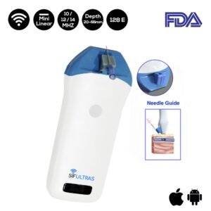
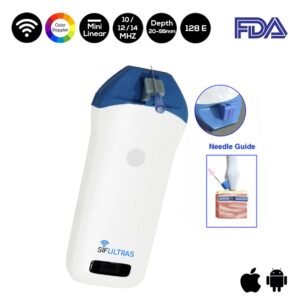
A mini linear transducer with high frequency offer real-time ultrasound guidance during PICC placement.
It has been proven that the mini linear ultrasound scanner reduces the incidence of tip malposition and insertional injuries.
The Ultrasound scanning modality is a valuable tool in vascular access. It does not only increases success rates but it also helps the practitioner avoid complications associated with lind needle sticks.
In addition the guidance that ultrasound offers, reduces the risks that patients might suffer, causing less vessel trauma than with blind insertion, gratifying the user the Ability to access deeper veins that are non-palpable and offer increased patient comfort and satisfaction. That is by letting you see veins clearly and steering you away from access sites compromised by such problems as vascular thrombosis or stenosis.
“When using ultrasound for venipuncture, position the ultrasound handheld probe (also called a transducer) on the skin to produce either transverse or sagittal images of veins. When you position the probe perpendicular to a vein, the vein appears as a circle on the ultrasound monitor screen; this is a transverse view. Placing the probe parallel to the vein produces a sagittal (longitudinal) view of the vein.
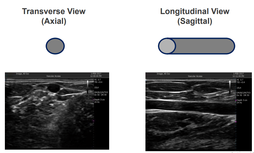
The transverse view is usually more helpful for guiding needle insertion. The sagittal view can help you follow a vein’s path, looking for valves, stenosis, and other abnormalities while moving the probe up the arm.
You’ll use your nondominant hand to hold the ultrasound probe and perform venipuncture with your dominant hand. To keep the insertion field sterile, place a sterile cover over the probe and use a sterile lubricant on the probe. Ask an assistant to make any necessary adjustments on the ultrasound machine while you work.” Source PICC Midline insertion workshop.
Like many other medical skills, using an ultrasound scanner requires practice, fine hand-eye coordination and a little imagination. While scanning, visualize the point and angle of entry required for the needle to enter the vein right under the probe. Imagine a two-dimensional pane of glass running through the patient’s arm; the needle should be positioned at such an angle that it would hit that pane right at the vein directly under the probe.
The ultrasound can also be used a Vessel assessment prior to insertion tool. As it assures vein is patent, determines appropriate size/depth of vessel, characteristics of pathway and blood flow in vessel, determines the location of surrounding anatomical structures. Further, it is an indispensable tool for the assessment of vessel after insertion • Determines continued patency of vein • Recognition of catheter related vessel thrombosis • Troubleshooting
PICC is inserted by : Anesthesiologist, Board certified radiologists trained in vascular interventional procedures, Qualified and specially trained radiology nurses, Radiology physician assistants or Radiology nurse practitioners …
Reference: PICC insertion procedure.
[launchpad_feedback]
Disclaimer: Although the information we provide is used by different doctors and medical staff to perform their procedures and clinical applications, the information contained in this article is for consideration only. SIFSOF is not responsible neither for the misuse of the device nor for the wrong or random generalizability of the device in all clinical applications or procedures mentioned in our articles. Users must have the proper training and skills to perform the procedure with each ultrasound scanner device.
The products mentioned in this article are only for sale to medical staff (doctors, nurses, certified practitioners, etc.) or to private users assisted by or under the supervision of a medical professional.