Ultrasound-guided Pleural Drainage PLEFF
Percutaneous pleural drainage PLEFF is the third most commonly performed procedure in the intensive care unit (ICU) after vascular catheterisation and tracheal intubation.
Ultrasound guidance allows the operator to increase the rate of success of the procedure and reduce its associated risks. Consequently, usage of the ultrasound guidance during pleural drainage has become mandatory.
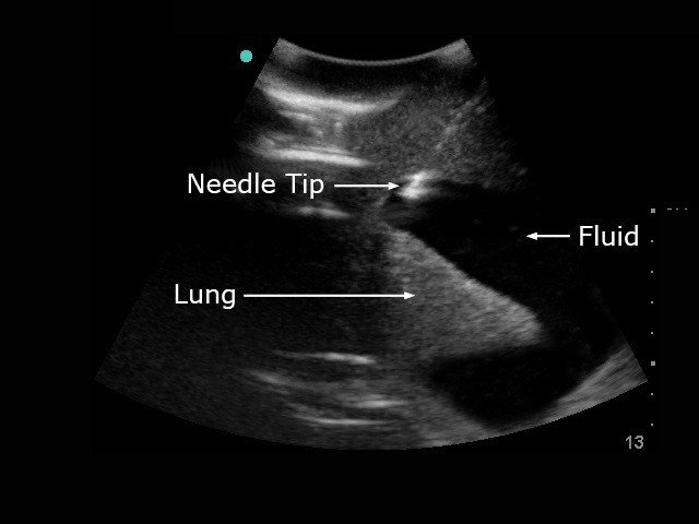
In addition to visualizing pleural effusion, thoracic ultrasound also helps clinicians to identify the best puncture site and to guide the drainage insertion procedure. Thoracic ultrasound is essential during these invasive manoeuvres to increase safety and decrease potential life-threatening complications.
Which ultrasound is best for pleural drainage?
Doppler ultrasound can be used to visualise intercostal vessels helping prevent vessel injury and ensure a procedure with a low risk of bleeding, even in patients with abnormal pre-procedural coagulation parameters.
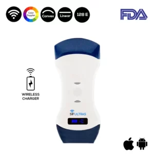
To Identify the best site for the puncture a low-frequency (3.5–5 MHz) convex proe of the SIFULTRAS-5.42 ultrasound transducer is recommended to identify the best puncture site evaluating these following steps. The probe should be used in the transverse position between two ribs
Then, the operator can Shift to the high-frequency (7–15 MHz) ultrasound transducer (linear probe of the SIFULTRAS-5.42). The probe should be used in the transverse position between two ribs to identify the upper and lower borders of the needle insertion area.
The puncture site and the needle trajectory must be carefully designed, with particular attention to the depth required to reach the pleural fluid and avoid lung injuries. In morbidly obese with limited rib palpation, linear probe can also be used to help to be perpendicular and over the upper rib edge with great safety.
The use of bedside ultrasound not only leads to an improvement in the diagnosis, but also allows the detection of the best puncture site and the fluid quantification of PLEFF. The positioning of percutaneous pleural drainage with ultrasound guidance increases the procedure’s success rate and safety
Ultrasound guidance allows the operator to better decide where to insert the pigtail. The best puncture site is, in fact, the place where the operator can visualise each anatomical structure (i.e., diaphragm, pleural, and organs) and where the operator can measure the maximum distance between visceral and parietal pleural (increasing the safety margin).
Doctors in most specialties will be exposed to patients requiring pleural drainage and need to be aware of safe techniques. Such as, cardiologist, emergency doctor, critical care specialist or anesthesiologist…

[launchpad_feedback]
Disclaimer: Although the information we provide is used by different doctors and medical staff to perform their procedures and clinical applications, the information contained in this article is for consideration only. SIFSOF is not responsible neither for the misuse of the device nor for the wrong or random generalizability of the device in all clinical applications or procedures mentioned in our articles. Users must have the proper training and skills to perform the procedure with each ultrasound scanner device.
The products mentioned in this article are only for sale to medical staff (doctors, nurses, certified practitioners, etc.) or to private users assisted by or under the supervision of a medical professional.


