
Ultrasound of Portal Vein Thrombosis
Portal vein thrombosis is blockage or narrowing of the portal vein (the blood vessel that brings blood to the liver from the intestines) by a blood clot. The the term portal venous thrombosis encompasses a broad spectrum of pathological conditions.
Portal vein thrombosis may be seen in a variety of clinical contexts, and when acute can be a life-threatening condition. It is a major cause of non-cirrhotic presinusoidal portal hypertension. Portal vein thrombus may be either bland and/or malignant (i.e. tumor thrombus), and it is a critical finding in liver transplant candidates, as it precludes transplantation.
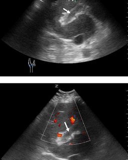
Acute thrombosis may be difficult to detect with grey-scale imaging alone, as the thrombus may be hypoechoic. With time, it becomes more echogenic and easier to identify. Color Doppler should be able to demonstrate absent flow in the portal vein and even to detect partial thrombosis, but attention to the Doppler gain and filters is necessary to avoid color overwrite of partial thrombosis.
Which ultrasound scanner is best for portal vein thrombosis diagnosis?
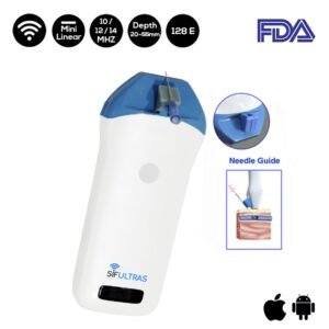
Color and spectral Doppler are used to evaluate flow characteristics in the portal and hepatic vessels. A high-frequency linear transducer SIFULTRAS-3.5 is used for PVT diagnosis.
Color Doppler is also useful to help evaluate for tumor thrombus, which will show internal color vascularity. Bland thrombus, in comparison, is avascular on color Doppler. The latter is the first-line imaging study for diagnosis of PVT; magnetic resonance angiography and CT angiography are valid alternatives.
It is much easier to obtain precise information on the abdominal vessels and their flow characteristics. As a result, PVT is diagnosed with increasing frequency. Important advances have also been made in the field of hereditary and acquired coagulation disorders, shedding new light on the potential causes of venous thrombosis.
In the past, thrombosis of the portal vessels was diagnosed with angiography or splenoportography. Today, the vessels of the splanchnic district can be accurately explored with noninvasive diagnostic methods like ultrasound or color Doppler ultrasound.
The sonographic diagnosis of portal vein thrombosis is based on demonstration of echogenic material that obstructs the lumen of the vessel and the complete or partial absence of flow in the portal vein or on the presence of collateral circuits that by-pass the obstructed vessel, the most typical form being the cavernoma, a tangle of tortuous vessels, irregular in caliber, that includes the vasa vasorum of the portal vein and the pericholecystic vessels.
If the obstruction is partial, color Doppler can reveal areas of the lumen that are patent and/or the presence of some flow downstream from the thrombus; the complete absence of flow should be confirmed with pulsed Doppler.
As cited in Portal vein thrombosis: Ultrasound imaging “the improvement of imaging procedures the number of patients with diagnosed portal vein thrombosis is increasingly growing. With a negative predictive value of 98% color Doppler ultrasound is considered as imaging modality of choice in detecting portal vein thrombosis.”
References: Portal vein thrombosis, Portal vein thrombosis: Ultrasound imaging.

[launchpad_feedback]
Disclaimer: Although the information we provide is used by different doctors and medical staff to perform their procedures and clinical applications, the information contained in this article is for consideration only. SIFSOF is not responsible neither for the misuse of the device nor for the wrong or random generalizability of the device in all clinical applications or procedures mentioned in our articles. Users must have the proper training and skills to perform the procedure with each ultrasound scanner device.
The products mentioned in this article are only for sale to medical staff (doctors, nurses, certified practitioners, etc.) or to private users assisted by or under the supervision of a medical professional.