
Intraoperative Vascular Ultrasound
A variety of intraoperative imaging and assessment techniques are available in modern operating rooms. However, there is no standardized method for assessing and evaluating vascular procedures.
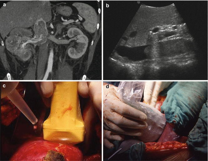
Ultrasound is a procedure that uses sound waves to see inside the body. An interoperative duplex ultrasound shows the surgeon the path and volume of blood flow before the surgery is completed. This exam is performed along with the surgical procedure.
The doctor may order an ultrasound to view and record the blood flow immediately during a surgical bypass procedure (repairing diseased arteries in the neck, kidneys or lower legs with healthy veins from the leg).
Which ultrasound scanner is used for intraoperative vascular ultrasound?
A linear 9.5 MHz ultrasound is used in vascular diagnosis intraoperatively. The vascular surgeon performs the ultrasound procedure following surgery but before the skin is closed. The SIFULTRAS-5.34 is placed in a sterile sleeve on the designated area.
The surgeon holds the transducer next to the blood vessel being evaluated. Sound waves bounce off the blood moving in the arteries and the tissue in the body. This creates echoes that are reflected back to the transducer.
Findings show that concomitant use of intraoperative vascular Doppler during microsurgical varicocelectomy allows a higher number of arterial branches preserved. Also more internal spermatic veins are likely to be ligated. This device should be considered attractive tool to improve surgical outcome and safety
The effect of Papaverine is reduced if the surgery is performed under epidural anesthesia, since basal flow is already increased. Obviously, blood flow values do not necessarily provide information about anatomical aberrations due to technical failure. In these cases intraoperative ultrasound imaging is performed in order to provide the surgeon with an anatomical evaluation as well
In addition, routine intraoperative duplex ultrasound examination of the carotid reconstruction allows early diagnosis and immediate correction of morphologic as well as hemodynamic lesions.
As the difference in transit times of the ultrasound beams is based only on moving elements in the blood vessel, the measurements are not influenced by the inner diameter of the vessel wall. This is especially relevant for atherosclerotic arteries.
This procedure is typically performed by a vascular surgeon.
References: Intraoperative Sonography During Carotid Endarterectomy,
Intraoperative Duplex Ultrasound.


[launchpad_feedback]
Disclaimer: Although the information we provide is used by different doctors and medical staff to perform their procedures and clinical applications, the information contained in this article is for consideration only. SIFSOF is not responsible neither for the misuse of the device nor for the wrong or random generalizability of the device in all clinical applications or procedures mentioned in our articles. Users must have the proper training and skills to perform the procedure with each ultrasound scanner device.
The products mentioned in this article are only for sale to medical staff (doctors, nurses, certified practitioners, etc.) or to private users assisted by or under the supervision of a medical professional.