Superficial and Deep Infrapatellar bursa
Infrapatellar bursitis causes pain and swelling at the front of the knee, just below the knee cap. It develops when there is irritation and inflammation of one of the small fluid-filled sacs in the knee.
The infrapatellar bursa is actually made up of two sacs:
* Superficial Infrapatellar Bursa: lies between the anterior subcutaneous tissues of the knee and the anterior surface of the patellar tendon
* Deep Infrapatellar Bursa: lies between the patellar tendon and the shin bone (tibia), it is characterised by inflammation of the bursal synovium and associated with the formation of an increased amount of fluid and collagen.
Ultrasound imaging helps the physicians evaluating the Infrapatellar region, in which it shows: distended deep infrapatellar bursa/cystic mass, internal septations, and heterogeneous soft tissue mass.
Which WiFi Ultrasound Scanner is the best in the assessment of Superficial and deep Infrapatellar bursa?
Orthopedists tend to use a high-frequency linear transducer in the evaluation of infrapatellar bursa. Such as the 5 to 12MHz Linear WiFi Ultrasound Scanner SIFULTRAS-9.53 that plays an important role in improving point-of-care, clinical diagnostic methods and efficiency of clinic diagnosis.
SIFULTRAS-9.53 guides the diagnosis and help delineate the presence of another knee bursitis, calcific tendonitis, tendinopathy, patellar tendonitis, or other knee pathology.
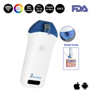
Physicians also might use the Color Doppler Mini Linear WiFi Ultrasound Scanner SIFULTRAS-3.51 that has the ability to provide real-time dynamic images of superficial soft tissues and is also used to carry out ultrasound-guided injections.
It comes with a needle guide holder. Hence, it can be directly set to the guide pin frame. Coupled with the software that can quickly locate the depth and diameter of puncture’s navigation. The latter allows the practitioner to visualize the needle in real-time as it enters the body and traverses to the desired location.
Ultrasonography is effective at diagnosing chronic fibrotic changes within the infrapatellar as well as inflammation associated with synovitis. Using a Color Doppler WiFi Ultrasound scanner helps indeed visualising the Inflammation within the fat pad.
References: Infrapatellar bursitis, Ultrasound-Guided Injection Technique for Deep Infrapatellar Bursitis Pain, Ultrasound-Guided Injection Technique for Superficial Infrapatellar Bursitis Pain, Deep infrapatellar bursitis,


