The Ultrasound-Guided Adductor Canal Block
The adductor canal (also known as Hunter’s canal or the subsartorial canal) is a narrow conical tunnel in the thigh.
It measures about 15cm from the apex of the femoral triangle to the adductor hiatus of the adductor Magnus. The canal is a passageway for structures that move between the anterior thigh and the posterior leg.
The Hunter’s canal is bordered by muscular structures:
· Anterior: Sartorius.
· Lateral: Vastus medialis.
· Posterior: Adductor Longus and adductor magnus.
The ultrasound-guided adductor canal block provides a reliable method for blocking the saphenous nerve. It is therefore a useful technique for postoperative analgesia following foot and ankle surgery.
The use of ultrasound (US) guidance has improved the success rates of the saphenous nerve blocks compared with field nerve blocks below the knee and blind trans-sartorial approaches. As with other nerve blocks, ultrasound guidance allows operators to visualize the nerve and can increase the efficacy and safety of the block.
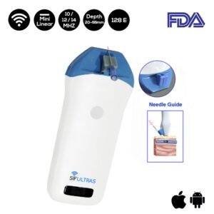
Moreover, the SIFULTRAS-3.5 Mini Linear Handheld Ultrasound Scanner has revolutionized the industry by allowing doctors to diagnose and treat patients more quickly and accurately. It is one of the most affordable and widely available imaging modalities.
Indeed, medical ultrasound scans are currently the most widely used imaging technique for adductor canal diagnosis and examinations in blocs.
The SIFULTRAS-3.5 Mini Linear Handheld Ultrasound Scanner has numerous advantages, ranging from reducing puncture complications to increasing patient satisfaction. SIFULTRAS-3.5 provides doctors with wireless Freedom as it connects to their tablet, smartphone, and it is iOS and Android compatible to give superior image quality.
Despite good intentions, even in the most experienced hands, blind (injections performed without imaging) injections are not 100% accurate and in some joints, accuracy is as low as 30%-40%. With ultrasound guidance, the accuracy of nearly every joint injection exceeds 90% and approaches 100% in many. Moreover, the device has a platform that is heavily software-based.
To conclude, a linear high-frequency ultrasound scanner is best for visualizing the adductor canal. SIFULTRAS-3.5 offers great resolution thanks to its higher frequency and advanced digital imaging technology .it is also useful in the emergency, clinic, plastic surgeries, EMS, anesthesia, MSK, joint injection, Acupuncture, abscesses, The insertion of the arterial lines, energy medicine, physiological medicine, naturopathy, and integrative medicine, for IV (intravenous injection), vein finding prior to injection, skincare, beauty clinics and offices outdoor/indoor, beauticians and vet inspections.
References: The adductor canal, Hunter’s Canal
ULTRASOUND-GUIDED ADDUCTOR CANAL BLOCK (SAPHENOUS NERVE BLOCK)

