Transvaginal Ultrasound Staging of Cervical Cancer
Cervical cancer is a common gynecologic malignancy worldwide. It is the fourth for both incidence and mortality. For cervical cancer, imaging and pathology assessments are incorporated in the revised 2018 Federation of Gynecology and Obstetrics (FIGO) staging system. Uses of imaging techniques for the pre-treatment work-up of cervical cancer have been increasing. Among imaging techniques for the evaluation of cervical cancer, ultrasound is cheaper, faster and widely available than other imaging techniques such as computed tomography (CT) or magnetic resonance imaging (MRI).
Advanced techniques in ultrasound, such as color Doppler, have improved the clinical application of ultrasound in cervical cancer. Ultrasound may provide highly accurate information on detecting tumor presence and evaluating local tumor extent if performed by ultrasound-trained gynecologists; the experience of readers is also critical for correct pretreatment staging and assessment of response to treatment. Sonographic images could be useful to predict response of neoadjuvant chemotherapy, radiotherapy, chemotherapy and concurrent chemoradiotherapy in patients with cervical cancer.
For cervical cancer, imaging and pathology assessments are incorporated in the revised 2018 Fédération internationale de gynécologie et d’obstétrique (FIGO) staging system. It is according to available resources for the use of the imaging modality to provide information on local or systemic spread, tumor size and nodal status. It was reported in a recent study that inclusion of imaging and surgical pathologic findings resulted in upward stage migration and mostly related to nodal and distant metastasis. Furthermore, incorporating the results of advanced imaging and surgical staging improved stratification of prognostic factors for relating survival outcomes compared to the FIGO 2009 staging system for most cases by a cohort of 1282 patients. Different sonographic appearances of adenocarcinoma (AC) and squamous cell cancer (SCC) of the cervix was reported. The researchers demonstrated isoechoic or hyperechoic (relative to the surrounding stroma) in AC and hypoechoic in SCC using transvaginal ultrasound (TVUS) . This could be helpful in the clinical evaluation of cervical tumors.
Another study at the Department of Gynecologic Oncology, National Cancer Institute, Milan, Italy has shown that 2D and 3D ultrasound showed similar moderate agreement with MRI. It’s proved that 2D and 3D ultrasound examinations are less costly and more readily available than MRI and should be considered in the preoperative work-up for cervical cancer.
Furthermore, Transvaginal Color Doppler ultrasound in cervical cancer allows non-invasive assessment of tumor angiogenesis. In Epstein et al study, Color Doppler signals were found in all cases of AC and 90% (18/20) of SCC cases. There was few detectable vascularization found in normal cervical tissue. Microvessel density correlated with survival for stage IB cervical cancer was assessed. Color Doppler findings associated with risk factors was also reported by a study involving neoangiogenesis measured in early cervical cancer cases.

Angiogenic parameters, including subjective assessment of pulsatility index (PI) and the amount of vessels within the tumor (scanty-moderate or abundant), were evaluated by transvaginal color Doppler ultrasound for 27 early stage cervical cancer patients. The risk factors (depth stromal invasion, parametrial and vaginal margin involvement, positive lymph nodes, lymph-vascular space involvement) were recorded. Tumors with abundant vascularization were significantly associated with parametrial involvement, lymph-vascular space involvement, pelvic lymph node metastases etc. Postoperative treatment was significantly more prevalent in patients with profuse vascularization.
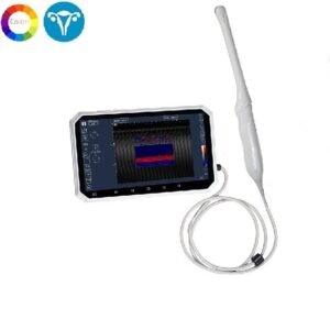
For this reason we highly recommend the Color Doppler Transvaginal USB Ultrasound Scanner 4-9 MHz SIFULTRAS-1.3 for the imaging and staging of cervical cancer.. It has a touch screen gesture and physical buttons for gain control, wavelet filter for Image processing, pseudo color, image smoothing, frame correlation and Accelerometer, P/L sensor, E-compass, and gyroscope. It measures depth, length and area. Equipped with a 5M pixels camera for telemedicine, Stereo FM radio and a 5000mAh battery, up to 3 hours of ultrasound continuous scans. This transvaginal ultrasound is also used by doctors to examine other female reproductive organs. This includes the uterus, urinary bladder, fallopian tubes, ovaries and vagina.
Cervical ultrasound imaging is performed by a qualified gynecologist whose is trained in ultrasound imaging*
Reference: Agreement of two-dimensional and three-dimensional transvaginal ultrasound with magnetic resonance imaging in assessment of parametrial infiltration in cervical cancer
Updated applications of Ultrasound in Uterine Cervical Cancer