
Traumatic Extremity Injuries
Choosing a surgical treatment strategy for patients with traumatic extremity injuries requires rapid detection, localization, and characterization of a possibly accompanying vascular injury.
Vascular injuries present a great challenge to emergency physicians because some vascular lesions may not be immediately identified with clinical evaluation and monitoring of vital signs.
Physical and sonographic examinations after extremity trauma have been found to be reliable means of detecting an occult arterial lesion.
Point-of-care ultrasound and Color flow Duplex Doppler ultrasound are widely available, cheaper, noninvasive, and faster to obtain. They can provide bedside valuable information for the identification of some vascular injuries allowing to an integrated management of the trauma patient, enriched by the use of ultrasound.
Which ultrasound scanner is best for traumatic extremity injuries assessment?
Vascular injuries of the extremities and neck must be investigated with high-frequency linear SIFULTRAS-5.42 transducers because a higher resolution and a lower penetration are commonly needed. There are some exceptions, such as patients with large hematomas and super obese patients, in which a convex SIFULTRAS-5.42 probe with higher penetration is recommended.
In the extremities and neck Point of care ultrasound is used for the diagnosis of fractures, hematomas, and the identification of foreign bodies. At the same time, in stable patients without hard signs of vascular damage, it could be of great help for a prompt bedside diagnosis, characterization, and monitoring of trauma-related vascular injuries.
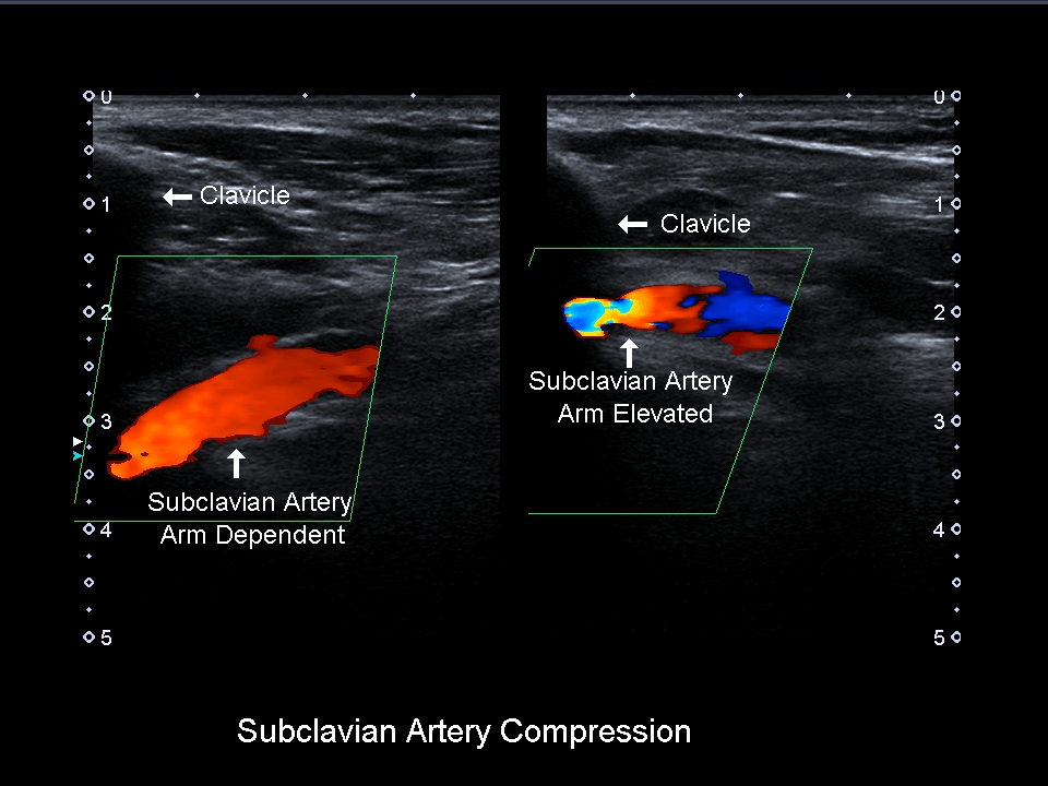
When evaluating vascular injuries, the region of the trauma must be explored, and transversal and longitudinal scans of the vessels should be performed with B-mode. Proximal and distal flows should be investigated with Color flow Duplex Doppler ultrasound. For comparative purposes, both limbs and both sides of the neck should be evaluated.
The pulsed wave Doppler analysis shows a continuous flow pattern with respiratory variations. Ultrasound can rapidly rule out vascular injuries after trauma at the bedside
Color Duplex Doppler evaluation will show the presence or absence of blood flow and the direction and velocity of the flow. In normal veins, the flow is continuous and spontaneous at rest and the velocity varies with respiration, increasing during inspiration, and decreasing during expiration or when a Valsalva maneuver is performed.
Point-of-care ultrasound and Color flow duplex Doppler ultrasound are widely available, noninvasive, sensible and specific techniques that can be used bedside, as a first approach for a prompt diagnosis and follow-up of trauma-related vascular injuries, and for an integrated management of trauma patient.
This procedure is done by an emergency doctor.


[launchpad_feedback]
Disclaimer: Although the information we provide is used by different doctors and medical staff to perform their procedures and clinical applications, the information contained in this article is for consideration only. SIFSOF is not responsible neither for the misuse of the device nor for the wrong or randomgeneralizability of the device in all clinical applications or procedures mentioned in our articles. Users must have the proper training and skills to perform the procedure with each vein finder device.
The products mentioned in this article are only for sale to medical staff (doctors, nurses, certified practitioners, etc.) or to private users assisted by or under the supervision of a medical professional.