
Ultrasonography for Sclerosing Adenosis
Sclerosing adenosis is a benign breast ailment that can develop as a result of aging. The lobules (milk-producing glands) and ducts (tubes that bring milk to the nipple) that make up the breasts are surrounded by glandular, fibrous, and fatty tissue.
When some of the lobules (milk-producing sacs) grow larger and contain more glands than usual, this is known as adenosis. Sclerosing adenosis is defined as an enlargement of the lobules with scar-like fibrous tissue. You or your doctor may notice a lump caused by adenosis.
Small breast lumps in a lobule characterize sclerosing adenosis. It may be painful, and you may see a bump on your chest.
Mammography may reveal sclerosing adenosis. It could be misinterpreted as breast cancer due to its deformed shape. To avoid similar misdiagnoses, an outstanding and crystal-clear ultrasound machine should be utilized.
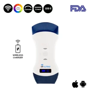
In this case, the SIFULTRAS-5.42 FDA high-resolution Color Double Head Wireless Ultrasound Scanner may be the finest option.
This one-of-a-kind portable ultrasound scanner features two heads, making it more practical and cost-effective than buying two single-headed probes, which may be required in this mammographic procedure.
The convex side of the color doppler transducer is utilized for in-depth inspections of interior body components such as these non-cancerous breast lumps, making it ideal for investigating fibroadenomas.
Surprisingly, this portable ultrasound scanner is designed specifically for radiologists to produce colored breast images and transfer them to their and their patients’ phones or tablet screens, allowing both parties to be fully aware of the problem and discuss the best treatment options in complete transparency.
The fact that this wireless ultrasound is compatible with both IOS and Android adds to its appeal. It’s small, light, and simple to use. As a result, radiologists are no longer restricted to their cabinets in order to complete such jobs.
The portable ultrasound: Color Double Head Wireless Ultrasound Scanner SIFULTRAS-5.42 FDA should be radiologists’ and Sclerosing Adenosis patients’ first choice because it is specifically built to view important internal organs such as the breast lobules in a very accurate method.
To summarize, Sclerosing Adenosis patients need not be concerned because they will receive accurate scan imagery as well as safer, faster, and more accurate tests. For this reason, we strongly suggest that Fibroadenomas patients use the handheld ultrasound scanner: SIFULTRAS-5.42.
Sclerosing lesions of the breast
Disclaimer: Although the information we provide is used by different doctors and medical staff to perform their procedures and clinical applications, the information contained in this article is for consideration only. SIFSOF is not responsible neither for the misuse of the device nor for the wrong or random generalizability of the device in all clinical applications or procedures mentioned in our articles. Users must have the proper training and skills to perform the procedure with each ultrasound scanner device.
The products mentioned in this article are only for sale to medical staff (doctors, nurses, certified practitioners, etc.) or to private users assisted by or under the supervision of a medical professional.