Ultrasound-Guided Hydatid Cyst Diagnosis
Hydatid disease (also known as echinococcosis or hydatidosis) is a potentially lethal disorder caused by cysts containing the larval stages of the Echinococcus granulosus.
The liver and lungs are the most common sites for hydatid cysts, but they can also appear in other organs, bones, and muscles. Cysts can grow to be 5–10 cm in diameter or more, and they can live for decades.
Infection with the larval stage of Echinococcus granulosus, a 2–7 millimeter long tapeworm found in dogs (definitive host), sheep, cattle, goats, and pigs, causes hydatid illness (intermediate hosts).
When hydatids develop in the liver, symptoms such as abdominal pain, nausea, and vomiting are prevalent. Chronic cough, chest pain, and shortness of breath are all symptoms of lung disease. Other symptoms vary depending on where the hydatid cysts are located and how much pressure they apply on the surrounding tissues.
Several imaging modalities, including ultrasound images, paired with case history, make it easy to make the diagnosis.
As a result, a professional and highly accurate scanning machine should be used to do all of these tasks flawlessly in order to provide accurate diagnosis and subsequent right treatment.
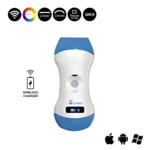
The Color Doppler 3 in 1 Wireless Ultrasound Scanner SIFULTRAS-3.31 is said to be specialists’ top recommendation because it met the ultrasonic parameters required for this specific scanning method.
This portable ultrasound scanner features a Convex 3.5/5MHz and a Linear 7.5/10MHz with an Electronic Array scanning mode. As a result, it’s ideal for recognizing the pseudocyst and, as a result, assuring an accurate hydatid disease diagnosis and, as a result, making the treatment procedure easier.
Furthermore, this wireless ultrasound scanner is widely used to monitor blood flow to vital organs such as the heart, kidney, liver, pancreas, and even muscles. This should imply that the ultrasound machine can produce exceptionally detailed scan images of those internal organs, allowing for a full diagnosis and evaluation of the problem.
The color doppler on the SIFULTRAS-3.31 mobile ultrasound scanner detects blood velocity in the affected area where the hydatid cyst is present. It’s worth noting that the probe’s head does not need to be replaced; instead, the software can be updated.
In conclusion, the SIFULTRAS-3.31 ultrasound machine is a fantastic portable ultrasound machine for any medical facility. Because of its basic interface, it does not require any additional training to utilize. It’s small, portable, and easy to use. But, most importantly, it’s great for people who have a hydatid cyst problem and are still waiting for a definitive diagnosis.
Reference: Hydatid Disease
Disclaimer: Although the information we provide is used by different doctors and medical staff to perform their procedures and clinical applications, the information contained in this article is for consideration only. SIFSOF is not responsible neither for the misuse of the device nor for the wrong or random generalizability of the device in all clinical applications or procedures mentioned in our articles. Users must have the proper training and skills to perform the procedure with each ultrasound scanner device.
The products mentioned in this article are only for sale to medical staff (doctors, nurses, certified practitioners, etc.) or to private users assisted by or under the supervision of a medical professional.

