Ultrasound-Guided Suprapatellar recess
Suprapatellar recess also known as the suprapatellar bursa or suprapatellar pouch is one of several bursae of the knee. It is located proximal to the knee joint, between the femoral and suprapatellar fat pads. As with all bursae, its purpose is to reduce friction between moving structures.
Inflammatory joint disorders usually affect the knee joint. Ultrasonography is equivalent to clinical assessment in evaluating the occurrence and location of knee joint effusion in some studies. In which ultrasonography detected soft tissue abnormality (suprapatellar bursitis, knee effusion, or Baker’s cyst) at 54/130 (42%) sites.
Which Ultrasound Scanner is the best for Suprapatellar recess assessment?
Using a high-frequency 5 to 12 MHz linear transducer is needed during the evaluation of suprapatellar bursa. For this reason, our medical research and development team always recommends the USB Linear 5-12MHz Ultrasound Scanner SIFULTRAS-9.53 to our orthopedist clients.
The SIFULTRAS-9.53 plays an important role in improving point-of-care, clinical diagnostic methods and efficiency of clinic diagnosis. With its sealed head and its USB connector, a steady ultrasound signal makes the signal transmission faster, and it provides a high-quality image that guides the physician to clear decisions during the assessment.
Indeed, the suprapatellar approach under ultrasound guidance avoids any tendons or bony or ligamentous structures and facilitates simple and accurate arthrocentesis for the provider.
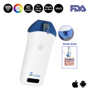
Physicians might also use the Color Doppler Mini Linear WiFi Ultrasound Scanner SIFULTRAS-3.51 for both the identification of the effusion and needle guidance for the arthrocentesis.
It helps to minimize attempts as well as improve procedural confidence in the hands of healthcare providers
The SIFULTRAS-3.51 comes with a needle guide holder. Hence, it can be directly set to the guide pin frame. Coupled with the software that can quickly locate the depth and diameter of puncture’s navigation.
Ultrasonography is effective in the identification of knee effusion as well as allow for a real-time visualized technique for joint aspiration. It can also be a useful adjunct in the evaluation of the patient with a swollen, painful knee.
References: Suprapatellar Bursa,


