Vitreous Hemorrhage Ocular Ultrasound Diagnosis
Vitreous hemorrhage (VH) is an important ophthalmic condition that may cause an abrupt decrease in visual acuity (VA) and it often occurs as a complication of an underlying disease. VH has an annual incidence of 7 to 15.4 cases per 100,000 persons, depending on the population studied. Some of the main causes of VH may be proliferative diabetic retinopathy (PDR), retinal vein occlusions (RVOs), ocular trauma, posterior vitreous detachment with or without a retinal tear, etc.
During the acute hemorrhagic event, blood passes through holes or apertures in the posterior hyaloid into the vitreous cortex, needing from weeks to months to clear from this location. A vitreous hemorrhage may result from proliferative retinopathy, the condition in which new, abnormal blood vessels grow on the surface of the retina. This is referred to as neovascularization. When not treated, these new blood vessels may continue to grow and spread through the vitreous onto the pupil area. This can increase ocular pressure (pressure within the eye) that presses on the optic nerve. Damage to the optic nerve is irreparable and can lead to vision loss. Bleeding from a vitreous hemorrhage can also cause scar tissue to form near the back of the eye. This can pull the retina away from the back lining of the eye, requiring additional treatment to keep the retina from detaching and permanently damaging vision.
Doctors will examine the patient’s eyes as well as review their medical history to determine the cause of the hemorrhage and recommend the appropriate treatment. To confirm the diagnosis, a series of diagnostic tests may be performed such as:
- Gonioscopy
- Dilated eye examination
- IOP
- Indirect ophthalmoscopy
- Slit-lamp examination
- B-scan
Nussenblatt´s standardization for vitreous opacities could be used as a system to clinically grade the opacities. In this scale, the clinical view through indirect ophthalmoscopy of the fundus is compared to a set of standard photographs with different degrees of vitreous haze. This scale is a straightforward way to categorize a VH, allowing the physician in day-to-day clinical practice to remember roughly the structures that need to be visible, in order to grade the hemorrhage without looking constantly at the reference images.
Although Nussenblatt’s grading scale has become the standard for more than 30 years, several problems with this system could be considered. First, it may have moderate interobserver agreement, as reported by Hornbeak et al. Secondly, as categorical variables, patients that fall in-between categories could yield to the subjective interpretation of each individual examiner and, therefore, may result in low agreement between observers; third, the scale does not allow an adequate measurement of sporadic or interventional improvement, regardless of the vitreous opacity. Therefore, a more objective and reproducible grading method could prove useful.
Quantification of vitreous hemorrhages (VH) termed minimum image gain (MIG) can be determined through ultrasound. Since its introduction into the ophthalmology field in 1956. Ocular ultrasound has become an invaluable tool that helps determine diagnoses and treatment decisions. All ultrasound systems allow adjustments in the amplification of the echo signals, in other words, the strength of the ultrasound beam. Changing the amplitude will modify the gain or sensitivity setting of the system. Gain is measured in a logarithmic scale in decibels (dB), which represent relative units of ultrasound intensity from the returning echo. Higher gain levels allow greater ability to display weaker echoes, such as vitreous opacities, whereas lower gain levels allow only stronger echoes, for example the sclera, to be displayed. Therefore, gain levels could prove useful as a measuring scale to determine the lowest signal intensity obtained from a specific structure (in the present case, the vitreous humor and VH).
The density of a specific tissue scanned by an ultrasound could be determined by knowing the acoustic impedance and the speed of sound in that tissue, which would imply having different software adaptations in the echography system. A more simple solution could be to modify the amplitude of the echo signals. Most (if not all) ocular ultrasound systems have the possibility to alter the gain or sensitivity to visualize a structure. With a lower gain, the amplitude of the ultrasound wave will not be strong enough and will attenuate as it goes through the tissue (in this case, the vitreous cavity). Higher sensitivity decreases attenuation, allowing the visualization of minute details. Decreasing the gain until no vitreous humor (or VH) is visualized (minimum gain) would mean that the specific density of the tissue (vitreous and hemorrhage) would be sufficient enough to attenuate the signal at that specific dB. VHs were demonstrated to have lower MIG measurements (52.8 dB) when compared with controls (77.97 dB). Because of the hemorrhage, the density of the vitreous is higher, and therefore, MIG is lower.
As per protocol: With the patient at dorsal decubitus position, a 10 MHz, B-scan ultrasound probe (with a depth of exploration of 20 to 60 mm, focus of 21 to 25 mm, axial resolution of 150 µm, and lateral resolution of 300 µm) is used to evaluate the temporal quadrant of the globe. A longitudinal image is obtained where the optic nerve head, macula, peripheral retina, and external rectus muscle could be visualized. Therefore, the 9 o’clock meridian is analyzed for right eyes and 3 o’clock for left eyes.
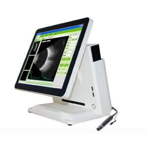
Based on the ocular ultrasound screening protocols for Vitreous hemorrhage we highly recommend the Ophthalmic Ultrasound Scanner SIFULTRAS-8.1. This ultrasound enables operators to easily image the anterior and posterior segments of the eye; providing important information not possible with clinical examination alone. Equipped with B-scan at Frequency range : 10MHz/20MHz (optional) ,Magnetic driven and noiseless, Real-Time magnification, 60 mm depth this device has proved to be an excelent choice for diagnosing Vitreous hemorrhage. It enhance the part of vitreous body and retina with a probe gain of 30dB-105dB perfectly suitable for grading vitreous hemorrage. Furthermore the SIFULTRAS-8.1 is equipped withan A-scan mode for anterior chamber depth, lens thickness, vitreous body length and total length measurements for cataract surgery to pick the right lense replacement and for diagnosis of tummors.
This procedure should be performed by a qualified ophthalmologist*
Reference: Vitreous Hemorrhage: Diagnosis and Treatment
Scale for Photographic Grading of Vitreous Haze in Uveitis