
Ultrasound-Assisted Percutaneous Needle Fasciotomy for Dupuytren’s Contracture
Dupuytren contracture is a debilitating disease that characteristically presents as a firm nodularity on the palmar surface of the hand with coalescing cords of soft tissue on the webs and digits.
The fascia is a layer of tissue that helps to anchor and stabilize the skin on the palm side of the hand. Without the fascia, the skin on the palm would be as loose and moveable as the skin on the back of the hand. In patients with Dupuytren’s disease, this palmar fascia slowly begins to thicken, then tighten.
Often, Dupuytren’s is first detected when lumps of tissue, or nodules, form under the skin in the palm. This may be followed by pitting on the surface of the palm as the diseased tissue pulls on the overlying skin.
As Dupuytren’s progresses, bands of fascia in the palm develop into thick cords that can tether one or more fingers into a bent position. This is called a “Dupuytren’s contracture.” Although the cords in the palm may look like tendons, the tendons are not involved in Dupuytren’s.
In many cases, a Dupuytren’s contracture progresses very slowly, over a period of years, and may remain mild enough such that no treatment is needed. In moderate or severe cases, however, the condition makes it difficult to straighten the involved digits. When this happens, treatment may be needed to help reduce the contracture and improve motion in the affected fingers. Typically, as a contracture worsens, the involvement of the fascia becomes more severe and treatment is less likely to result in a full correction.
In mild to moderate cases of Dupuytren contractures Percutaneous needle fasciotomy (PNF) is the common treatment solution. For most patients PNF is performed in a regular outpatient office after local disinfection of the skin. The coalescing cord is punctured at it’s thinnest part. Through extention of the joint the punctures on the coalescing cord will rupture releasing the tension on the finger.
Although percutaneous needle fasciotomy for Dupuytren’s contracture is a simple, inexpensive procedure, it is a blind procedure with risks including injury to nerves, arteries, and tendons.
In 2015 a study has been conducted on a novel technique using ultrasound as an adjunct to percutaneous fasciotomy for Dupuytren’s contracture. Generally, patients have no postoperative restrictions other than to avoid submerging their hands for 48 hours. To date, the authors have noted, in 66 cases, no permanent complete nerve dysfunction following needle aponeurotomy using ultrasound assistance. The study has concluded that Ultrasound mapping of the digital neurovascular structures can be successfully used as an adjunct to help prevent these neurovascular complications.
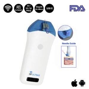
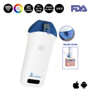
For this application we highly recommend the Color Doppler Mini Linear WiFi Ultrasound Scanner SIFULTRAS-3.51 and the Mini Linear Handheld WiFi Ultrasound Scanner 10-12-14 MHz, SIFULTRAS-3.5. These two portable scanners comes with a needle guide holder. Hence, it can be directly set to the guide pin frame. Coupled with the software that can quickly locate the depth and diameter of the puncture’s navigation. It allows the practitioner to visualize the needle in real time as it enters the body and traverses to the desired location.
This operation is done by an orthopedic specialist.
Reference:
+Management of Dupuytren contracture with ultrasound-guided lidocaine injection and needle aponeurotomy coupled with osteopathic manipulative treatment
+Ultrasound-Assisted Percutaneous Needle Fasciotomy for Dupuytren’s Contracture
+Modified dynamic high-frequency ultrasound-guided needle aponeurotomy for Dupuytren’s contracture
[launchpad_feedback]