Ultrasound-guided Core-needle Biopsy of Breast lesions
Ultrasound-guided core-needle biopsy on the breast is a reliable alternative to surgical biopsy for a histological diagnosis. Percutaneous biopsy is less invasive than surgery. It can be performed quickly, does not deform the breast, causes minimal scarring, complications (haematoma and infection) are infrequently encountered (less than one case in 1,000), fewer surgeries are needed for patients who undergo percutaneous biopsies and consequently the cost of diagnosis is lower.
There are two main objectives of percutaneous biopsy techniques: first, achieving the maximum degree of accuracy and second, offering as much information as possible about the tumour (type, grade, invasion, hormonal receptors, HER-2 NEU, etc.). To achieve these objectives, the percutaneous biopsy devices have evolved, from fine-needle aspiration cytology towards core-needle biopsy (CNB) and later vacuum-assisted biopsy (VAB).
Nowadays, ultrasound-guided core-needle breast biopsy has become the first choice for performing a percutaneous biopsy for most lesions seen on ultrasound. Virtually any breast lesion that is clearly seen on ultrasound can be sampled under ultrasound guidance. Prior to the performance of any ultrasound-guided percutaneous procedure, the lesion should be evaluated
completely with a diagnostic ultrasound in accordance with the ACR Practice Parameter for the Performance of a
Breast Ultrasound Examination and assessed by a physician qualified to interpret the examination . Findings on other imaging modalities (such as mammography or MRI) or on clinical examination should
be correlated with those seen by ultrasound before the interventional procedure is undertaken.
Advantages:
When a lesion can be visualized sonographically, ultrasound guidance is generally preferred over other modalities because it is often faster, allows real-time imaging of the sampling process, and is more comfortable for the patient.
Ultrasound requires neither ionizing radiation, unlike mammographic modalities, nor intravenous contrast, unlike
MRI. Using ultrasound guidance when appropriate also has cost advantages because procedure times tend to be
shorter and ultrasound equipment is less expensive and often more readily available than the other modalities.
Additionally, both mammographic and MRI-guided biopsies generally require vacuum-assisted devices, whereas
ultrasound biopsies can usually be performed with either lower-cost spring-loaded large-core biopsy devices or,
when appropriate, vacuum-assisted devices.
Furthermore intra-tumoral blood-flow analysis well correlates with aggressiveness and histological grade of the mass, so a preoperative assessment using Color Doppler may give preliminary prognostic informations useful for therapeutic planning. Color Doppler ultrasound may be valuable also in assessing the efficacy of neoadjuvant chemotherapy and in particular of antiangiogenesis treatments.
Disatvantages
The main disadvantage of ultrasound core-needle biopsy is the limitation of performing a biopsy on lesions that arer not seen on ultrasound. Most clustered microcalcifications, especially if they are not inside a mass, cannot be identified on ultrasound. However, high-resolution transducers can demonstrate some clustered microcalcifications even in the absence of a mass. Although most ultrasound CNB procedures are easy to perform, in some special situations (deeply located lesions, patients with implants, axillary lesions, etc.) a high level of experience is needed to get reliable results.
The use of high-frequency (10- to 12-MHz) probes, adjustments in the dynamic range and postprocessing grey scales, as well as correct focus, are important to improve the visibility of breast lesions. After localising the lesion with ultrasound, the procedure is performed in an outpatient setting, using the free-hand technique: one hand holds the probe and the other hand holds the needle. One of the main advantages of ultrasound-guided core-needle biopsy is the full control of the needle position in real time, allowing for corrections in the needle direction.
As a general rule, the shortest route from the skin to the lesion should be used. A vertical approach would be the best, but it is not possible under ultrasound guidance. However, an oblique approach, as parallel to the chest wall as possible, should be used. This is the way to avoid pneumothorax, the worst complication of this technique. This approach also enables the best visualisation of the needle, because even large-gauge needles are difficult to visualise if a steep angle is used because of less reflective echoes. However, when the needle is parallel to the probe, the number of needle-generated reflected echoes that are perpendicular to the ultrasound beam is maximised, so the needle can be identified. This horizontal approach can be used to perform a biopsy for cutaneous lesions.
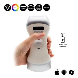
The 3 in 1 Color Doppler Wireless Ultrasound Scanner SIFULTRAS-3.32 with it’s high-frequency linear probe, particularly at 7.5–10 MHz makes for a great guide to core-needle biopsy of breast lesions . Featuring Elements: 192 this ultrasound provides an ulta high resolution imaging. Thanks to it’s smaller footprint it aids in evaluation of soft tissues leading to an accurate core-needle biopsy. Equipped with color doppler system the SIFULTRAS-3.32 could provide preliminary prognostic informations useful for therapeutic planning through intra-tumoral blood-flow analysis which correlates with aggressiveness and histological grade of the mass as previously covered. This scanner is also equipped with a convex probe making for is a highly versatile device that serves various medical practitioners and fields
Ultrasound-guided percutaneous breast interventional procedures should be performed by physicians who meet
qualifications outlined in the ACR Practice Parameter for the Performance of a Breast Ultrasound Examination *
References: Ultrasound-guided core-needle biopsy of breast lesions
Has color Doppler a role in the evaluation of mammary lesions?