
Cardiomegaly Diagnosis Ultrasound
A weak heart muscle, coronary artery disease, heart valve abnormalities, or irregular heart rhythms are among medical conditions that may lead to cardiomegaly. The heart may enlarge due to the thickening of the cardiac muscle or dilatation of one of the heart’s chambers. A cardiac enlargement may be either transient or permanent, depending on the underlying cause.
An enlarged heart may be the result of conditions that cause the heart to beat too rapidly or that cause damage to the heart muscle. The heart may enlarge and weaken for unknown causes. Commonly known as “idiopathic cardiomegaly,” this condition occurs for no apparent reason. Many things may lead to a bloated heart, including congenital cardiac defects, heart attack damage, and arrhythmia.
Some persons with enlarged hearts show no outward symptoms. These symptoms may also be present in others: Weakness in breathing, Rhythm disturbances in the heart (arrhythmia), Excessive Bloating (edema)
Ultrasound imaging may be used to diagnose heart enlargement, which, if untreated, might cause heart failure, blood clots, or cardiac arrest. Because of this, an ultrasound is recommended for anybody experiencing breathlessness, cardiac arrhythmia, or edema.
Ultrasound imaging has obvious diagnostic applications. One major problem is that not all scanners now in use are of a high enough quality to be considered professional. In order to achieve clear scan images and a precise diagnosis, both doctors and patients need to exercise utmost care while choosing a device.
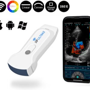
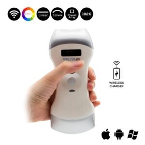
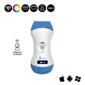
The Color Doppler 3 in 1 Wireless Ultrasound Scanner SIFULTRAS-3.31 is usually recommended by cardiologists in this situation. The most promising technique for identifying enlarged hearts is wireless ultrasound. First, it has a cardiac probe that greatly aids the doctor in determining the heart’s health by measuring the blood’s flow direction and velocity.
Therefore, this technology gives excellent information on the development of the problem and the potential therapy or medications that should be given, as well as clear scan results of the damaged region of the heart, which can be seen instantly by the doctor or surgeon.
This unique gadget solely detects volume and % vascularity when paired with 2D Mode, as has been discussed, and it has excellent Color picture quality, accurate scan findings is cost-Effective, tiny and light, simple to carry and use, and is ideal for evaluating the severely sick. The SIFULTRAS-3.31 is one of the best ultrasound scanning machines thanks to its cutting-edge capabilities, which allow for rapid and precise diagnosis of the Enlarged Heart problem and subsequent effective treatment.
Reference: Cardiomegaly
Disclaimer: Although the information we provide is used by different doctors and medical staff to perform their procedures and clinical applications, the information contained in this article is for consideration only. SIFSOF is not responsible neither for the misuse of the device nor for the wrong or random generalizability of the device in all clinical applications or procedures mentioned in our articles. Users must have the proper training and skills to perform the procedure with each ultrasound scanner device.
The products mentioned in this article are only for sale to medical staff (doctors, nurses, certified practitioners, etc.) or to private users assisted by or under the supervision of a medical professional.