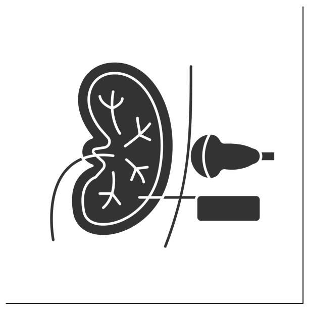
Speed and Direction of Blood Flow through the Vessel Ultrasound
Using the Doppler ultrasound, it is possible to measure motions within the body. Imaging of blood vessels using the Doppler mode, is a test that uses standard ultrasound methods to make a picture of a blood vessel and the organs around it. The portable ultrasound scanner App turns the Doppler sounds