
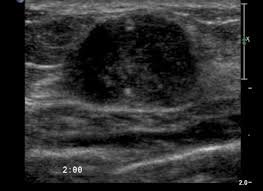
Breast Masses
Breast mass is a cause of great concern. High frequency, high-resolution ultrasonography (USG) helps in its assessment.
This is represented in women with dense breast tissue where USG is useful in discovering small breast cancers that are not seen on mammography.
Which ultrasound scanner is used for breast imaging?
SIFULTRAS-5.39 ultrasound scanner with high-frequency technology, particularly with 7.5–10 MHz probes, has brought out a totally new facet in USG breast masses imaging. It is also a medical masterpiece not only in sonography of the breast but also in the Thyroid, Vascular, Nerve, MSK (Musculoskeletal).
Ultrasound carries definitely no risk for any patient and can be carried out easily in comparison to computed tomography or magnetic resonance imaging (MRI).
By sonographic images it is possible to distinguish between benign lesions and malignant lesions of the breast. Also, the Ultrasound is essential for diagnosing the breast lump type and determining the best treatment.
Under the USG, masses were classified into five types: cystic, complex, solid, calcification, and unclassifiable. Therefore, Ultrasound is essential for the diagnosis and classification of breast lumps after breast augmentation by autologous fat grafting, and can be used to select the appropriate treatment in accordance with the condition of the lumps.
The procedure is performed by Breast surgeons, oncologists, pathologists…


[launchpad_feedback]
Although the information we provide is used but doctors, radiologists, medical staff to perform their procedures, clinical applications, the Information contained in this article is for consideration only. We can’t be responsible for misuse of the device nor for the device suitability with each clinical application or procedure mentioned in this article.
Doctors, radiologists or medical staff must have the proper training and skills to perform the procedure with each ultrasound scanner device. Toggle panel: Post Options