
Mullerian abnormalities
Mullerian abnormalities happen when müllerian channels grow abnormally, which can damage the growth of the entire generative system, including the fallopian tubes, uterus, cervix, and upper two-thirds of the vagina.
Which ultrasound scanner is used for Mullerian anomalies assessment?
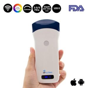
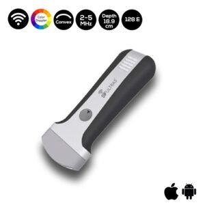
3D Ultrasound scanner with a convex probe with low frequency SIFULTRAS-5.21 and/or endocavitary probe is best used to visualize the Müllerian anomalies of the uterine.
The capacity to accurately recognize the type of an anomaly becomes hard as pregnancy advances. It is noninvasive, relatively cheap and appropriate, as most pregnant women go through ultrasound testing routinely.
However, early in pregnancy three-dimensional ultrasound is used to assess both the uterine contour and cavity. The use of cervical length ultrasound to stratify patients at greatest risk of preterm delivery is routinely used in other high-risk obstetric populations and may prove beneficial in women with uterine anomalies.
(source : Mullerian abnormalities ).
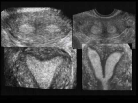
On 3D ultrasound, the connection of the cavity with the fundus is obvious, allowing accurate diagnosis thanks to the contribution of the C‐plane (coronal) that is impossible to acquire in most cases on 2D ultrasound yet is essential to the diagnosis of these anomalies.
Not to mention the ability to observe uterine malformations with less extreme forms (arcuate, septate and bicornuate uterus) two cavities with further details.
Additionally, 3D ultrasound allows us to make measurements such as the length and thickness of the septum, calculate the volume of the cavity and study the vascularization, which can affect fertility prognosis, thus aiding the choice of treatment.
The success of 3D ultrasound in the diagnosis of uterine malformations is well recorded in the medical field. (
Source: ( Three‐dimensional ultrasound in the diagnosis of Müllerian duct anomalies ).
This procedure is done by an Obstetrician, gynecologist ..

[launchpad_feedback]
Although the information we provide is used but doctors, radiologists, medical staff to perform their procedures, clinical applications, the Information contained in this article is for consideration only. We can’t be responsible for misuse of the device nor for the device suitability with each clinical application or procedure mentioned in this article.
Doctors, radiologists or medical staff must have the proper training and skills to perform the procedure with each ultrasound scanner device.