
Ultrasound-guided Caudal Injection
A Caudal Injection is an injection into the lowest portion of the epidural space, this can help reduce lower back and leg pain caused by sciatica, herniated discs, bone spurs or other back problems.
This operation consists of inserting a thin needle into the patient’s back (above the tailbone), injecting dye to confirm if the medication is administered into the caudal space, after that, inducing a mixture of anesthetics and steroid (for long term relief).
Which ultrasound scanner is used for caudal injections?
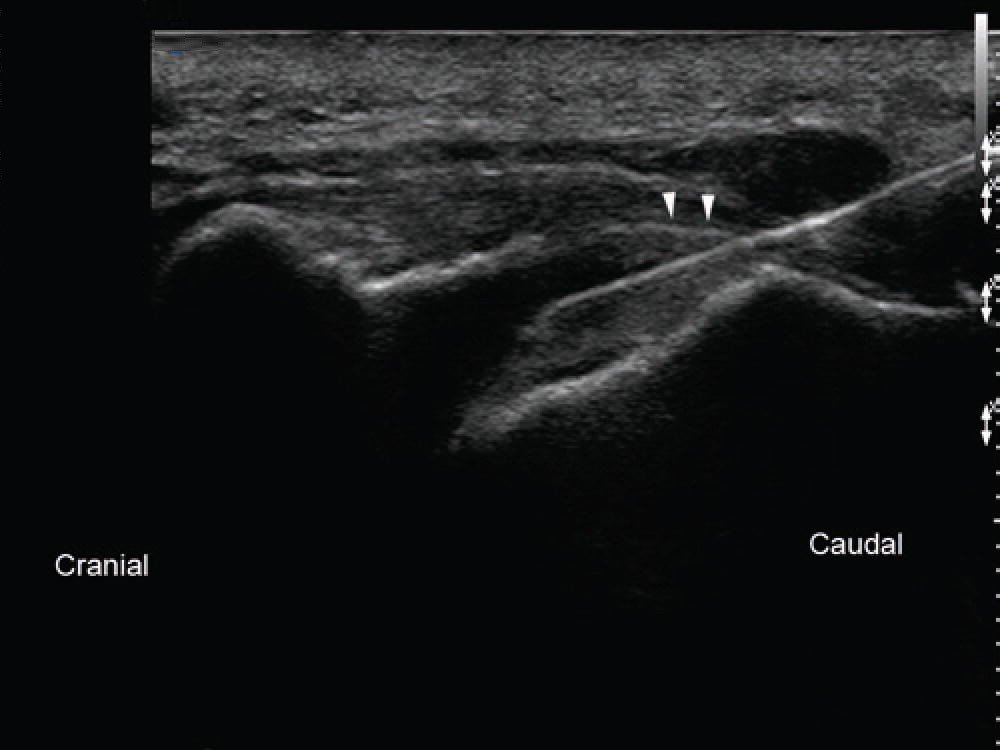
This is where the Ultrasound Scanner comes handy, using the Ultrasound Scanner SIFULTRAS-5.31, it is placed transversely at the midline to obtain a transverse sonographic view of the sacral hiatus.
Then, the transducer is rotated 90 degrees to rest between the two cornua and to obtain the longitudinal sonographic view of the sacral hiatus. After that, the doctor proceeds to advancing the needle between the two cornua to the sacral hiatus and then into the caudal epidural space.
Ultrasound-guided caudal injection technique has been shown to have 100% accuracy in correct needle placement into the sacral canal for subsequent epidural injection, and is the most accurate and safe way to perform this procedure.
Caudal injection are performed by pain management specialist, nurses, anesthesiologists..


[launchpad_feedback]
Although the information we provide is used but doctors, radiologists, medical staff to perform their procedures, clinical applications, the Information contained in this article is for consideration only. We can’t be responsible for misuse of the device nor for the device suitability with each clinical application or procedure mentioned in this article.
Doctors, radiologists or medical staff must have the proper training and skills to perform the procedure with each ultrasound scanner device.