
FAST: Focused Assessment with Sonography in Trauma
The Focused Assessment with Sonography in Trauma (FAST) is a point-of-care ultrasound examination performed at the time of presentation of a trauma patient.
It is an ultrasound protocol developed to identify intraperitoneal free fluid (assumed to be hemoperitoneum in the context of trauma) allowing for an immediate transfer to theater, CT or other.
Which ultrasound scanner for Focused Assessment with Sonography in Trauma?
The 2 MHz to 5 MHz curvilinear (or abdominal) probe is used for the eFAST exam to eliminate delays when switching between transducers. However, the phased array (or cardiac) probe is effective as well, particularly with parasternal windows. Likewise, the 5 MHz to 12 MHz linear (or vascular) probe is ideal for assessing for pleural sliding. Making the SIFULTRAS-3.31 the device of choice in FAST exams.
The use of ultrasound device (US) in trauma has expanded to identifying a variety of traumatic injuries: hemoperitoneum, pneumothorax, hemothorax, hemopericardium with or without tamponade, traumatic hypovolemia, and even rib, nose, and other fractures! The convex head helps you to quickly detect free intraabdominal fluid and the cardiac head shelps you to quickly detect any cardiac complications that may occur.
The extended FAST, or E-FAST, expands the examination to assess for pneumothorax. The primary indications for performing a FAST are blunt or penetrating trauma, trauma in pregnancy, or hypotension of unclear etiology.
A FAST using the wireless portable color doppler double head ultrasound scanner helps determine which patients require emergent laparotomy and which can be monitored or await slower, more definitive studies.
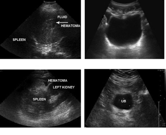
The philosophy behind the FAST examination is that fluid will pool in the most dependent areas.
As a result, the FAST examination includes 3 views that can detect pooled blood (red lines) and one to evaluate the heart: the hepatorenal recess, the perisplenic view, the subxiphoid pericardial window, and the suprapubic window.
The linear 7.5 – 10 MHz probe is effective as well, particularly with parasternal windows. Likewise, it is ideal for assessing for pleural sliding.
The implementation of point of care ultrasound has significantly impacted the evaluation and treatment of patients.
Ultraspund have considerable advantages, including their bedside availability, ease of use, and reproducibility. Furthermore, it is non-invasive, employs no radiation or contrast agents, and is inexpensive.
The use of ultrasound to detect intraperitoneal fluid was first described its use decreases time to surgical intervention, patient length of stay, and rates of Computed tomography (CT) and Diagnostic peritoneal lavage (DPL).
Focused assessment with sonography in trauma (commonly abbreviated as FAST) is a rapid bedside ultrasound examination performed by surgeons, emergency physicians, and certain paramedics as a screening test for blood around the heart (pericardial effusion) or abdominal organs (hemoperitoneum) after trauma

[launchpad_feedback]
Although the information we provide is used but doctors, radiologists, medical staff to perform their procedures, clinical applications, the Information contained in this article is for consideration only. We can’t be responsible for misuse of the device nor for the device suitability with each clinical application or procedure mentioned in this article.
Doctors, radiologists or medical staff must have the proper training and skills to perform the procedure with each ultrasound scanner device.