Identifying Perforators in Reconstructive Surgery
Pedicled perforator flaps allow the surgeon to relocate local tissue and facilitate a simple reorganization, which enables an optimal cosmetic and functional reconstructive outcome. They provide a fast and simple, single-stage solution and offer an alternative to microsurgery or skin graft.
Handheld Doppler and color Doppler ultrasonography (CDU) have been shown to be useful to identify perforators and aid in the planning of flap reconstructions
Which ultrasound scanner is best for identifying perforators in reconstructive surgery?
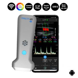
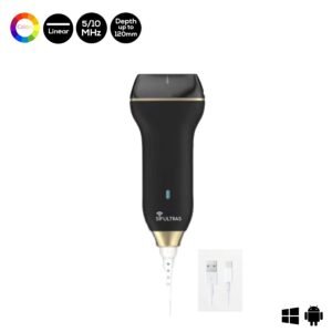
To map perforator locations an 8MHz Doppler device is advised the SIFULTRAS-5.34. CDU provides additional visual information about available soft tissue, vessel flow patterns, vessel course through the soft tissue as well as perforator size and location.
Doppler sonography is useful for locating the position of individual perforating vessels, making it much easier to find them during the operation
The individual perforating vessels have a high degree of anatomical variation, therefore it is desirable to conduct a careful examination of them before undertaking a perforator flap operation.
Locating the vessels beforehand makes performing the operative procedure much easier.
The CDU examination could easily identify the locations, numbers and the courses of the cutaneous perforators and help in the useful assessment of vascularity to confirm the existence and location of appropriate perforators for the design of the SDMC flap.
Using this CDU examination, difficulties owing to the anatomical variation of perforators are easily overcome, simplifying flap harvest.
Identifying perforatores are done by Plastic and reconstructive surgeons.
References: The Value of Preoperative Doppler Sonography for Planning Free Perforator Flaps, Color Doppler ultrasonography targeted reconstruction using pedicled perforator flaps, Color Doppler ultrasound assessment for identifying perforator arteries of the second dorsal metacarpal flap.

[launchpad_feedback]
Although the information we provide is used but doctors, radiologists, medical staff to perform their procedures, clinical applications, the Information contained in this article is for consideration only. We can’t be responsible for misuse of the device nor for the device suitability with each clinical application or procedure mentioned in this article.
Doctors, radiologists or medical staff must have the proper training and skills to perform the procedure with each ultrasound scanner device.


