
Ultrasound-guided Lateral Femoral Cutaneous Nerve
The lateral femoral cutaneous nerve (LFCN) divides into several branches innervating the lateral and anterior aspects of the thigh. The variable anatomy of the lateral femoral cutaneous nerve makes it challenging to perform an effective landmark-based block.
LFCN has received much attention because of its association with meralgia paresthetica. Knowledge of its anatomical variations is also crucial for preventing nerve injury during the insertion of needles into the ASIS, the harvesting of the anterior iliac crest bone grafts, and other surgical procedures. LFCN grafts can also be used to repair facial nerve injuries and soft-tissue defects.
Research prove that the rate of successful anesthesia has only been approximately 40% based on the use of anatomical landmarks. Ultrasound guidance, however, allows for more accurate needle insertion into the appropriate fascial plane through which the LFCN passes.
Which ultrasound scanner is best for Lateral Femoral Cutaneous Nerve?
A linear high-frequency ultrasound scanner is best for visualizing the LFCN. SIFULTRAS-3.5 offer great resolution thanks to its higher frequency, allows the practitioner to identify the LFCN if the intermuscular space between the tensor fasciae latae muscle and the sartorius is used as an initial sonographic landmark.
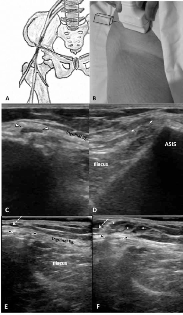
The LFCN typically is visualized between the tensor fasciae latae muscle (TFLM) and the sartorius muscle (SaM), 1–2 cm medial and inferior to the anterior superior iliac spine (ASIS) and 0.5–1.0 cm deep to the skin surface.
Ultrasound (US) imaging of the LFCN yields an oval hypoechoic small structure with a hyperechoic rim that can be easily seen in the hypoechoic background. The LFCN can be traced proximally, as it runs from the lateral to the medial edge of the superficial fascia of the SaM.
The lateral edge of the SaM is a useful landmark, and as such, it can be relied on throughout the procedure. The posterior branch of the LFCN may sometimes be seen across the anterior margin of the TFLM.
There are several promising studies about the ability of ultrasound in the evaluation of the LFCN, the use of ultrasound guidance in regional anesthesia for blocking the LFCN and the use of ultrasound in nerve conduction studies of the LFCN.
Reference: Ultrasound of the lateral femoral cutaneous nerve in asymptomatic adults, Ultrasound-Guided Lateral Femoral Cutaneous Nerve Block.
[launchpad_feedback]
Disclaimer: Although the information we provide is used by different doctors and medical staff to perform their procedures and clinical applications, the information contained in this article is for consideration only. SIFSOF is not responsible neither for the misuse of the device nor for the wrong or random generalizability of the device in all clinical applications or procedures mentioned in our articles. Users must have the proper training and skills to perform the procedure with each ultrasound scanner device.
The products mentioned in this article are only for sale to medical staff (doctors, nurses, certified practitioners, etc.) or to private users assisted by or under the supervision of a medical professional.