
Iterscalene Plexus Nerve Blocks (PNB)
The interscalene approach to brachial Plexus Nerve Blocks results in reliable anesthesia of the shoulder and upper arm. The supraclavicular branches of the cervical plexus, supplying the skin over the acromion and clavicle, are also blocked due to the proximal and superficial spread of local anesthetic. The inferior trunk (C8-T1) is usually spared unless the injection occurs at a more distal level of the brachial plexus.
Ultrasound guidance allows for visualization of the spread of the local anesthetic and additional injections around the brachial plexus if needed to ensure an adequate spread of local anesthetic, improving block success. Consequently, reducing local anesthetic volume required to accomplish the block.
Which ultrasound scanner for the interscalene plexus nerve block?
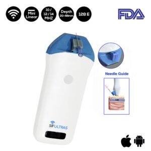
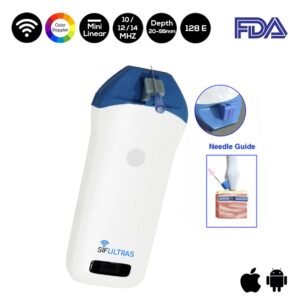
An ultrasound machine with a linear transducer (10–14 MHz) SIFULTRAS-3.5 , is needed for this procedure. The brachial plexus is typically visualized at a depth of 1–3 cm.
Scanning usually begins just below the level of the cricoid cartilage and medial to the sternocleidomastoid muscle with the goal of identifying the carotid artery. Once the artery has been identified, the transducer is moved slightly laterally across the neck.
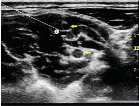
The goal is to identify the anterior and middle scalene muscles and the elements of the brachial plexus that is located between them. It is recommended to use the color Doppler to identify vascular structures and avoid them. The needle is then inserted in-plane toward the brachial plexus, typically in a lateral-to-medial direction
The brachial plexus at the interscalene level is seen lateral to the carotid artery and internal jugular vein, between the anterior and middle scalene muscles. Further, An ultrasound-guided technique can also decrease the incidence of hemidiaphragmetic paresis.
This PNB clinical application may be performed by an Anesthesiologist.
[launchpad_feedback]
Although the information we provide is used but doctors, radiologists, medical staff to perform their procedures, clinical applications, the Information contained in this article is for consideration only. We can’t be responsible for misuse of the device nor for the device suitability with each clinical application or procedure mentioned in this article.
Doctors, radiologists or medical staff must have the proper training and skills to perform the procedure with each ultrasound scanner device.