Ultrasound Scanner for Polycystic Ovarian Syndrome Assessment
Polycystic Ovarian Syndrome (PCOS) is a hormonal condition that women can get during their reproductive age. It is a disorder involving infrequent, irregular, or prolonged menstrual periods.
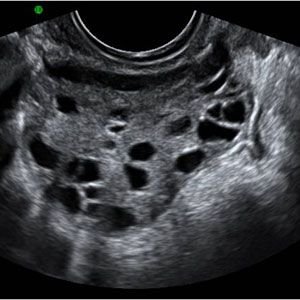
During the PCOS the ovaries may develop numerous small collections of fluid ( follicles) and fail to regularly produce eggs. Consequently lead to infertility, hypertension, hyperlipidemia, type 2 diabetes, coronary artery disease and cerebral vascular disease.
Moreover, women with PCOS are also at greater risk for endometrial hyperplasia and carcinoma as well as breast and ovarian cancers.
According to The Journal of Clinical Endocrinology & Metabolism, PCOS affects approximately 6.6 % of women in the United States, making it the most common endocrine abnormality in women of reproductive age.
Indeed, Color Doppler Ultrasound Scanner is an imaging modality that has the potential to improve the sensitivity and specificity of ultrasound without adding much to the practical management of the syndrome.
Besides, when it comes to its role in the PCOS identification, Ultrasound is helpful to predict fertility outcome in patients undergoing treatment.
Which Ultrasound Scanner is the best for Polycystic Ovarian Syndrome Assessment?
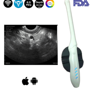
Ultrasound is considered the best standard in the diagnosis of polycystic ovaries. Thus, SIFSOF ‘s medical research and development team tends to suggest the Convex and Transvaginal Color Double Head Wi-Fi Ultrasound Scanner SIFULTRAS-5.43 to our gynecologist clients.
The Transvaginal probe is used to examine the uterus, fallopian tubes, ovaries, cervix, and vagina. While the Convex side of the Doppler is used for an in-depth examination of the internal parts of the body.
Therefore, the Double-Head SIFULTRAS-5.43 is suitable for more than one application at the same time. In which, the low-frequency convex probe 3.5 to 5MHz meets the doctor’s needs so that it can go deep from 100 to 200 mm to monitor and provide an accurate diagnosis.
In addition, the Transvaginal Probe 6.5 MHz is effective for detecting the appearance of polycystic ovaries in women with PCOS. Due to the optimal visualization it provides, of the internal structure of the ovary.
To sum up, Ultrasound Scanner has many reported advantages, including lower cost, wider availability, non-invasiveness, and lack of ionizing radiation. Most importantly, using Ultrasound for the assessment of the PCOS is helpful in which it helps first in its discovery and diagnosis and last but not least, it is effective during the treatment and the follow up stages.
References: Ultrasound Assessment of the polycystic ovary, Ultrasound Assessment of the Polycystic Ovaries,