Speed and Direction of Blood Flow through the Vessel Ultrasound
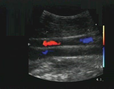
Using the Doppler ultrasound, it is possible to measure motions within the body. Imaging of blood vessels using the Doppler mode, is a test that uses standard ultrasound methods to make a picture of a blood vessel and the organs around it. The portable ultrasound scanner App turns the Doppler sounds into a graph. This graph helps to show the speed and direction of blood flow through the blood vessel.
Doppler ultrasound works by measuring sound waves that are reflected from moving objects, such as red blood cells. This is known as the Doppler effect.
Gives an image of the blood vessel that represents the speed and direction of blood flow through the vessel?
Color Doppler SIFULTRAS-5.34 This type of Doppler uses a feature in the App to change sound waves into different colors. These colors show the speed and direction of blood flow in real time.
Power Doppler, SIFULTRAS-3.21 a newer type of color Doppler. It can provide more detail of blood flow than standard color Doppler. But it cannot show the direction of blood flow, which can be important in some cases.
Spectral Doppler SIFULTRAS-9.52 This test shows blood flow information on a graph, rather than color pictures. It can help show how much of a blood vessel is blocked.
Duplex Doppler SIFULTRAS-5.17 This test uses standard ultrasound to take images of blood vessels and organs. Then a computer turns the images into a graph, as in spectral Doppler.
Continuous wave Doppler. SIFULTRAS-5.37 In this test, sound waves are sent and received continuously. It allows for more accurate measurement of blood that flows at faster speeds.
Medical Doppler ultrasound is usually utilized in the clinical adjusting to evaluate and estimate blood flow in both the major (large) and the minor (tiny) vessels of the body. The normal and abnormal sign waveforms can be shown by spectral Doppler technique.
Doppler ultrasound tests are used to help health care providers find out if you have a condition that is reducing or blocking the blood flow. It may also be used to help diagnose certain heart diseases. The test is most often used to:
Check heart function. It is often done along with an electrocardiogram, a test that measures electrical signals in the heart.
Look for blockages in blood flow. Blocked blood flow in the legs can cause a condition called deep vein thrombosis (DVT).
Check for blood vessel damage and for defects in the structure of the heart.
Look for narrowing of blood vessels. Peripheral arterial carotid artery stenosis.
Monitor blood flow after surgery.
Check for normal blood flow in a pregnant woman and her unborn baby.
A Doppler ultrasound test uses reflected sound waves to see how blood flows through a blood vessel. It helps doctors assess the blood flow through major arteries and veins, such as those of the arms, legs, and neck.
Doctor specialties who typically have to measure blood flow velocity are: Cardiologist, cardiovascular surgeon or a Hematologist
Reference : Doppler Ultrasound
[launchpad_feedback]
Although the information we provide is used but doctors, radiologists, medical staff to perform their procedures, clinical applications, the Information contained in this article is for consideration only. We can’t be responsible for misuse of the device nor for the device suitability with each clinical application or procedure mentioned in this article.
Doctors, radiologists or medical staff must have the proper training and skills to perform the procedure with each ultrasound scanner device.


