The use of Color Doppler Ultrasound with Transvaginal probe
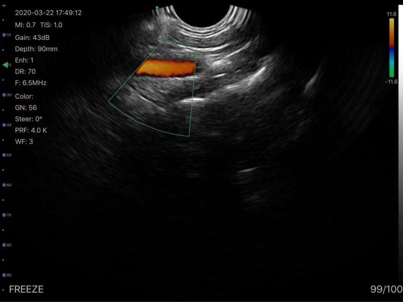
Color Doppler is a type of Doppler Ultrasound that converts the Doppler measurements into an array of colors. This color visualization is combined with a standard ultrasound picture of a blood vessel to show the speed and direction of blood flow through the vessel.

Transvaginal ultrasound has been the foremost imaging technique to assess the female pelvis. The physician can receive images in real-time and perform the pelvic examination simultaneously.
Indeed, Color Doppler Ultrasound with a Transvaginal probe is needed for the evaluation of gynecologic diseases. For this reason, our gynecologist clients opt to choose the double-head Color Doppler Convex and Transvaginal Ultrasound Scanner SIFULTRAS-5.43.
The Transvaginal probe is used to examine the uterus, fallopian tubes, ovaries, cervix, and vagina. While the Convex side of the Doppler is used for an in-depth examination of the internal parts of the body.
Gynecologists use Color Doppler Transvaginal Wireless Ultrasound Scanner in various cases such as an abnormal pelvic, unexplained vaginal bleeding, pelvic pain, an ectopic pregnancy, Infertility, a check for cysts or uterine fibroids, and verification that an IUD is placed properly.
Besides, the use of transvaginal color Doppler and color Doppler energy allows the assessment of ovarian endometrioma vascularity.
A study on the use of Transvaginal Color Doppler to assess blood circulation in pelvic vessels have shown that Blood flow was successfully displayed by color Doppler in the external and internal iliac arteries, and the uterine arteries.
Moreover, In early pregnancy blood flow in the umbilical artery could be visualized by color Doppler starting from the 6th week and flow in the aorta from the 8th week. Flow in the trophoblasts was observed with an overall success rate of 59% and successfully demonstrated in 1 out of 2 cases of ectopic pregnancy.
To sum up, the Color Doppler Transvaginal probe helps the physician to study and observe the female reproductive system. It is used to show the speed and direction of blood flow in certain pelvic organs.
References: Transvaginal Sonography, Vaginal Ultrasonography, Transvaginal Doppler Color Flow Mapping,


