Ultrasonography and Pseudocysts
A pseudocyst is a common consequence of pancreatitis, both acute and chronic. Pseudocysts are not actual cysts since the lining of the cyst is made up of scar tissue created by surrounding organs when pancreatic fluid leaks during an acute injury, rather than true pancreatic cells.
Pseudocysts can be caused by the following factors:
- Injury to the pancreas.
- Infection of the pancreas.
- Pancreatic cancer is a type of cancer that affects the pancreas.
- Calcium levels in the blood are too high.
- Blood fat levels are extremely high (cholesterol)
- Medicines can cause pancreatic damage.
- Autoimmune illnesses are a type of autoimmune disease.
- Conditions that affect the pancreas and run in your family, such as cystic fibrosis.
Patients with pancreatic cysts may not exhibit any symptoms. Pancreatic cysts are frequently identified by chance during abdominal imaging examinations that the patient is having for another cause. Pancreatic cysts are sometimes detected as a result of pain or after a pancreatitis crisis.
When symptoms or signs do appear, they may include:
- Consistent abdominal pain that may spread to your back
- Jaundice is a type of jaundice.
- Nausea and vomiting are common side effects.
- Weight loss that hasn’t been explained
- Diarrhoea that hasn’t been explained
Therapeuticendoscopists (gastroenterologists with extensive training in endoscopic operations) use modern diagnostic techniques such as endoscopic ultrasound (EUS) to visualize the cyst and help determine its type. Fine needle aspiration of the cyst is also possible with endoscopic ultrasonography. This allows tumor markers and cytology to be used to assist diagnose the cyst’s type, which is crucial for designing an effective treatment plan.
To provide correct diagnosis and subsequent proper treatment, a professional and clear scanning machine should be employed to execute all of these jobs perfectly.
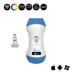
Because it satisfied the ultrasonic criteria required for this specific scanning process, the Color Doppler 3 in 1 Wireless Ultrasound Scanner SIFULTRAS-3.31 is stated to be specialists’ top suggestion.
With an Electronic Array scanning mode, this portable ultrasound scanner has a Convex 3.5/5MHz and a Linear 7.5/10MHz. As a result, it’s great for precisely detecting the pseudocyst and so ensuring an accurate spleen diagnosis, easing the treatment process.
This wireless ultrasound scanner is frequently used to monitor blood flow to important organs like the heart, kidney, liver, pancreas, and, of course, the spleen. This should imply that this ultrasound equipment is capable of producing extremely detailed scan images of those interior organs, ensuring a thorough diagnosis and evaluation of the issue at hand.
The SIFULTRAS-3.31 mobile ultrasound scanner also has a color doppler for detecting blood velocity in the damaged spleen area. It’s worth mentioning that the probe’s head does not need to be replaced because the software can be updated instead.
In summary, the SIFULTRAS-3.31 ultrasound machine is an excellent portable ultrasound machine for any medical establishment. It does not require any extra training to use due to its simple interface. It’s light, portable, and simple to operate. But, most importantly, it is ideal for individuals suffering from Pseudocysts who are looking for a precise diagnosis.
Reference: Pseudocysts
Disclaimer: Although the information we provide is used by different doctors and medical staff to perform their procedures and clinical applications, the information contained in this article is for consideration only. SIFSOF is not responsible neither for the misuse of the device nor for the wrong or random generalizability of the device in all clinical applications or procedures mentioned in our articles. Users must have the proper training and skills to perform the procedure with each ultrasound scanner device.
The products mentioned in this article are only for sale to medical staff (doctors, nurses, certified practitioners, etc.) or to private users assisted by or under the supervision of a medical professional.

