
Ultrasound Diagnosis of Renal Column Hypertrophy
Renal column hypertrophy represent the extension of renal cortical tissue which separates the pyramids, and as such are normal structures. They become of radiographic importance when they are unusually enlarged and may be mistaken for a renal mass.
Renal column hypertrophy is easily misdiagnosed as an intrarenal space occupying lesion, which needs to be distinguished from small renal space occupying lesions such as cysts and small tumors.
Renal cysts show a complete capsule, with no echo, and enhanced posterior echo. Small renal tumors often show a complete border or capsule. The echo is often lower or higher than the renal cortical echo.
Renal column hypertrophy has its clear features on the B ultrasound image, so it is generally not necessary to do other imaging examinations.
The internal echo is often uneven, sometimes corresponding. Dynamic observation of the sonogram changes, and the corresponding clinical symptoms may appear. The space-occupying lesions have a spherical sense.
Renal column is formed by the fusion of normal glomeruli. Under normal circumstances, the inner end is generally flush with the tip of the renal pyramid. When the renal column is abnormally enlarged, it protrudes into the renal sinus, and a localized low echo area can be seen in the collection system, which can be round and easily misdiagnosed as a renal mass occupying lesion.
Some hypertrophic kidney columns protrude into the renal collecting system in a long column shape, almost matching the medial cortex and renal hilum, but close observation shows that the hypoechoic column is not connected to the contralateral cortex and renal hilum, and the collecting systems at both ends are not isolated, some cut planes can still be shown as a whole, and would not distinguish the two renal hilum.
A hypoechoic nodule is seen in the renal sinus, which is evenly distributed, echoed with the cortex, and connected to it, causing the renal sinus to split on one side. When the renal column hypertrophy is prominent, it can cause the renal sinus to split into a sound image like double renal pelvis.
Which ultrasound is used for renal column hypertrophy?
In the adult patient, a curved array transducer with center frequencies of 3–6 MHz SIFULTRAS-5.21 is used, while the pediatric patient should be examined with a linear array transducer with higher center frequencies. Artifacts of the lowest ribs always shadow the upper poles of the kidneys.
The hypertrophic kidney column can be round, long cylindrical and tapered, the outer side is connected to the cortex, the remaining boundary is clear and smooth, and some hypertrophic kidney column can show a complete low echo on any surface.
The “occupied” clump is caused by the strong echo of the renal sinus sags toward the sides of the hypertrophic kidney column, but the clump has no sense of sphere. Renal column hypertrophy occurs in the lateral cortex of the kidney, and is more common in the upper middle part.
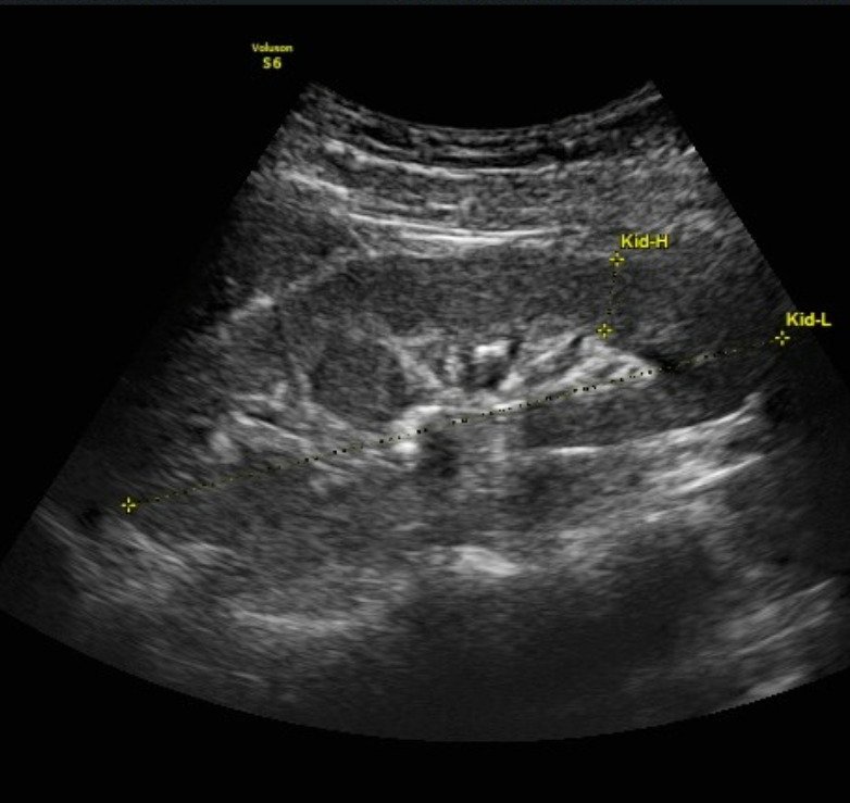
The right kidney is more common than the left kidney, and it is also seen in both kidneys. The diameter of the hypertrophic kidney column is generally ≤3 cm. The maximum diameter of the hypertrophic renal column in this group is 2.8 cm.


[launchpad_feedback]
Disclaimer: Although the information we provide is used by different doctors and medical staff to perform their procedures and clinical applications, the information contained in this article is for consideration only. SIFSOF is not responsible neither for the misuse of the device nor for the wrong or random generalizability of the device in all clinical applications or procedures mentioned in our articles. Users must have the proper training and skills to perform the procedure with each ultrasound scanner device.
The products mentioned in this article are only for sale to medical staff (doctors, nurses, certified practitioners, etc.) or to private users assisted by or under the supervision of a medical professional.